

Auditory Cortical Plasticity in Patients with Single-Sided Deafness Before and After Cochlear Implantation
- PMID: 38253897
- PMCID: PMC10907329
- DOI: 10.1007/s10162-024-00928-3
Auditory Cortical Plasticity in Patients with Single-Sided Deafness Before and After Cochlear Implantation
Abstract
Purpose: This study investigated neuroplastic changes induced by postlingual single-sided deafness (SSD) and the effects of a cochlear implantation for the deaf ear. Neural processing of acoustic signals from the normal hearing ear to the brain was studied before and after implantation using a positron emission tomography (PET)/CT scanner.
Methods: Eight patients with postlingual SSD received a cochlear implant (CI) in a prospective clinical trial. Dynamic imaging was performed in a PET/CT scanner using radioactively labeled water ([15O]H2O) to localize changes in the regional cerebral blood flow (rCBF) with and without an auditory task of logatomes containing speech-like elements without meaningful context. The normal hearing ear was stimulated before implantation and after the use of the cochlear implant for at least 8 months (mean 13.5, range 8.1-26.6). Eight age- and gender-matched subjects with normal hearing on both sides served as healthy control subjects (HCS).
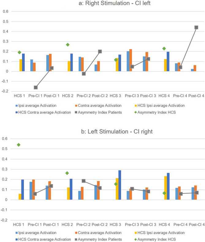
Results: When the normal hearing ear of SSD patients was stimulated before CI implantation, the [15O]H2O-PET showed a more symmetrical rCBF in the auditory regions of both hemispheres in comparison to the HCS. The use of CI increased the asymmetry index (AI) in six of eight patients indicating an increase of activity of the contralateral hemisphere. Non-parametric statistics revealed a significant difference in the AI between patients before CI implantation and HCS (p < .01), which disappeared after CI implantation (p = .195).
Conclusion: The functional neuroimaging data showed a tendency towards normalization of neuronal activity after CI implantation, which supports the effectiveness of CI in SSD patients.
Trial registration: ClinicalTrials.gov Identifier: NCT01749592, December 13, 2012.
Keywords: CI; Cerebral blood flow; H215O; SSD; Unilateral hearing loss.
© 2024. The Author(s).
Conflict of interest statement
The authors declare no competing interests.
Figures


References
MeSH terms
Associated data
Grants and funding
LinkOut - more resources
Full Text Sources
Medical
