. 2024 Jan 22;10(1):8.
doi: 10.1186/s40942-024-00529-5.

Serous maculopathy with absence of retinal pigment epithelium (SMARPE) associated with large drusen

Affiliations

Serous maculopathy with absence of retinal pigment epithelium (SMARPE) associated with large drusen

Luiz H Lima et al. Int J Retina Vitreous. .

Abstract

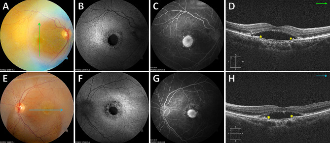
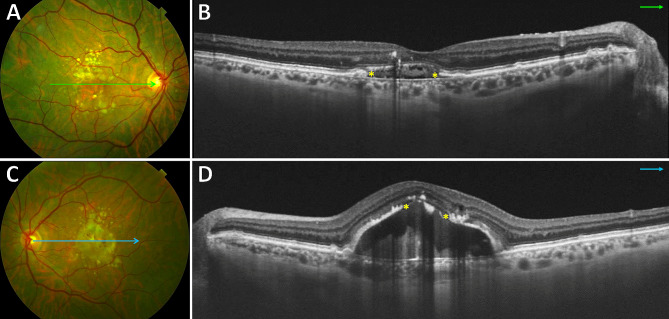
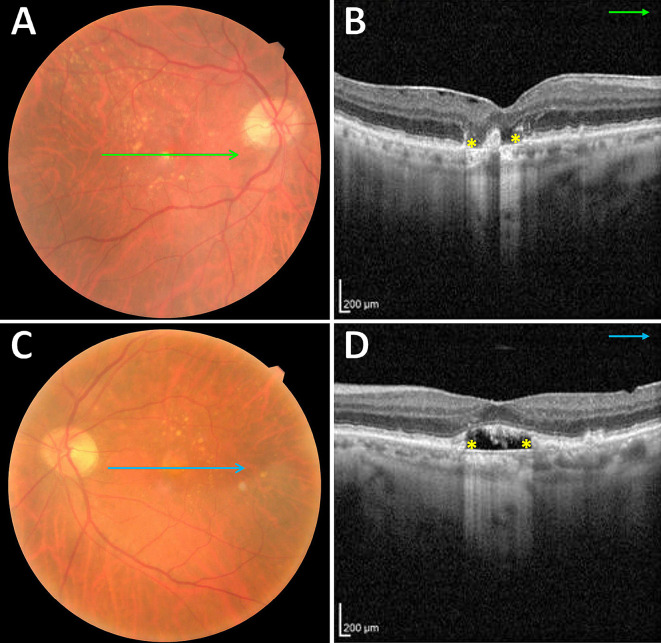
Purpose: To describe the association of serous maculopathy with absence of retinal pigment epithelium (SMARPE) and large drusen in patients with non-neovascular age-related macular degeneration (AMD).

Methods: A retrospective study of ophthalmic examination and multimodal imaging data of individuals with SMARPE and large drusen observed over a period of 12-month was accomplished. SMARPE was defined as subretinal accumulation of fluid within the macular area due to retinal pigment epithelium (RPE) aperture. Large drusen were identified by the presence of sub-RPE deposits using multimodal imaging analysis (color fundus photography, fundus autofluorescence, and spectral-domain optical coherence tomography).

Results: Twelve eyes of 7 white patients with a mean age of 77 years were observed to have SMARPE associated with large drusen. The median visual acuity was 20/100. Bilateral SMARPE lesions were observed in 71% of study patients. All SMARPE lesions were hypoautofluorescent, located in the subretinal space between the RPE and the ellipsoid zone, and presented as complete or incomplete RPE apertures associated with subretinal fluid. The SMARPE in this study had coincident multimodal imaging features as the SMARPE described in other reports in the literature.

Conclusions: Bilateral SMARPE can occur in association with typical AMD large drusen. Anomalisms resulting in drusen biogenesis or mechanisms that act alongside to these may be related to SMARPE development.

Keywords: Age-related macular degeneration; Large drusen; Multimodal imaging; Serous maculopathy with absence of retinal pigment epithelium (SMARPE).


Conflict of interest statement

GM is a scientific consultant to SJJ Solutions and Molecular Partners, and has been part of advisory boards of Bayer, Molecular Partners, and Roche. The other authors declare that they have no competing interests.

Figures

Fig. 1
Fig. 1
Multimodal imaging in a 75-year-old woman (Case # 1) presenting with bilateral serous maculopathy with absence of retinal pigment epithelium (SMARPE). Visual acuity was 20/150 in the right eye and 20/200 in the left eye. Color fundus photograph of both eyes (A and E) shows several typical age-related macular degeneration (AMD) large drusen and retinal pigment epithelium (RPE) atrophy within the macular area, with correspondent hypoautofluorescence on fundus autofluorescence (FAF) (B and F), and hyperfluorescence due to window defect on fluorescein angiography (FA) (C and G). Cross-sectional spectral-domain optical coherence tomography (SD-OCT) of both eyes (D and H) depicts the incomplete type (asterisks) of SMARPE (there is part of the RPE between the SRF edges) in the macular area. An intact ellipsoid zone (EZ) and external limiting membrane (ELM) overlying the RPE defect are observed in both eyes
Fig. 2
Fig. 2
Multimodal imaging in a 69-year-old man (Case #3) with bilateral SMARPE. Visual acuity was 20/80 in the right eye and 20/60 in the left eye (A and C). Color fundus photograph of both eyes demonstrates RPE atrophy surrounded by an agglomerate of AMD large drusen within the macular area. (B and D). Cross-sectional SD-OCT shows complete and incomplete types (asterisks) of SMARPE in the right and left eyes, respectively
Fig. 3
Fig. 3
Multimodal imaging in an 80-year-old man (Case #7) with unilateral SMARPE. Visual acuity was 20/80 in the right eye and 20/50 in the left eye. (A and C). Color fundus photograph of both eyes demonstrates several typical AMD large drusen within the macular area. (B and D). Cross-sectional SD-OCT shows a complete RPE and outer retinal atrophy (cRORA) (asterisks) in the right eye, and a complete type (asterisks) of SMARPE in the left eye

References

    1. van Dijk EHC, Boon CJF. Serous business: delineating the broad spectrum of diseases with subretinal fluid in the macula. Prog Retin Eye Res. 2021;84:100955. doi: 10.1016/j.preteyeres.2021.100955. - DOI - PubMed
    1. van Dijk EHC, Diederen RM, Boon CJF. Serous maculopathy with absence of retinal pigment epithelium (SMARPE) Acta Ophthalmol. 2022;100:583–8. doi: 10.1111/aos.14995. - DOI - PubMed
    1. Querques G, Capuano V, Costanzo E, et al. Retinal pigment epithelium aperture: a previously unreported finding in the evolution of avascular pigment epithelium detachment. Retina. 2016;36(Suppl 1):65–S72. doi: 10.1097/IAE.0000000000001231. - DOI - PubMed
    1. Goldstein BG, Pavan PR. Blow-outs’ in the retinal pigment epithelium. Br J Ophthalmol. 1987;71:676–81. doi: 10.1136/bjo.71.9.676. - DOI - PMC - PubMed
    1. Giannakaki-Zimmermann H, Querques G, Munch IC, et al. Atypical retinal pigment epithelial defects with retained photoreceptor layers: a so far disregarded finding in age related macular degeneration. BMC Ophthalmol. 2017;17:67. doi: 10.1186/s12886-017-0452-0. - DOI - PMC - PubMed

LinkOut - more resources