. 2024 Jan 4;14(1):68.
doi: 10.3390/biom14010068.

Effect of Topical Programmed Death-Ligand1 on Corneal Epithelium in Dry Eye Mouse

Affiliations

Effect of Topical Programmed Death-Ligand1 on Corneal Epithelium in Dry Eye Mouse

Ko Eun Lee et al. Biomolecules. .

Abstract

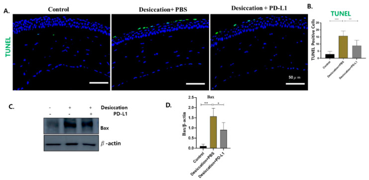
Dry eye disease (DED) is a growing health concern that impacts millions of individuals every year, and is associated with corneal injury, excessive oxidative stress and inflammation. Current therapeutic strategies, including artificial tears and anti-inflammatory agents, are unable to achieve a permanent clinical cure due to their temporary nature or adverse side effects. Therefore, here, we investigated the effectiveness of the topical administration of programmed death-ligand 1 (PD-L1) in the mouse model of DED. The model was generated in C57BL/6 mice by excising the extra orbital lacrimal gland and causing desiccation stress with scopolamine injections. Subsequently, either phosphate-buffered saline (3 µL/eye) or PD-L1 (0.5 µg/mL) was topically administered for 10 days. Tear volume was evaluated with phenol red thread, and corneal fluorescein staining was observed to quantify the corneal epithelial defect. Corneas were collected for histological analysis, and the expression levels of inflammatory signaling proteins such as CD4, CD3e, IL-17, IL-1β, pIkB-α, pNF-kB and pERK1/2 were assessed through immunofluorescence and Western blot techniques. Our results demonstrate that desiccating stress-induced corneal epithelial defect and tear secretion were significantly improved by topical PD-L1 and could reduce corneal CD4+ T cell infiltration, inflammation and apoptosis in a DED mouse model by downregulating IL-17 production and ERK1/2-NFkB pathways.

Keywords: corneal epithelial; desiccation stress; dry eye disease; programmed death-ligand 1.


Conflict of interest statement

The authors declare no conflict of interest.

Figures

Figure 1
Figure 1
Comparisons of the PD-L1 for DED mouse corneal epithelial recovery. (A) Fluorescein-stained mice corneas in control, desiccation+PBS and dessication+PD-L1 group were assessed by slit-lamp microscopy on days 0, 5 and 10 (B) Corneal fluorescein staining in control, desiccation+PBS and desiccation+PD-L1 group was presented with a bar graph. (C) Tear volume rate (mm wetted in 15 s) in a DED mouse model over 10 days. (* p < 0.05; ** p < 0.01; significant difference by one-way ANOVA).
Figure 2
Figure 2
Changes in the corneal morphological alterations in control, desiccation+PBS and desiccation+PD-L1 group. H&E staining showed that superficial CECs in the control group were well arranged and tightly attached and the corneal surface was smooth. The corneal epithelial layers were disrupted in the desiccation+PBS, and were improved in the desiccation+PD-L1 group (n = 6, day 10).
Figure 3
Figure 3
(A) Immunofluorescence staining showing the expression of (i) PD-L1, (ii) CD4, (iii) CD3e and (iv) IL-17 localized in the corneas in the control, desiccation+PBS and desiccation+PD-L1 group. (BE) Changes in the percentage of PD-L1, CD4, CD3e and IL-17 positive cells in the cornea. Immunopositivity for PD-L1 is shown in higher-power fields. Immunopositivity was counted in low-power fields and calculated as relative to the total number of DAPI-positive cells. (F,G) Western blot showing the expression of CD4 and IL-17 in the cornea. (H,I) Relative expression ratio of CD4 and IL-17 to β-actin. In (BE,H,I), data are presented with bar graph (n = 6, day 10, * p < 0.05; ** p < 0.01; *** p < 0.001 significant difference by one-way ANOVA). Original images of (F,G) can be found in Supplementary Materials.
Figure 4
Figure 4
(A) Immunofluorescence staining showing the expression of IL-1β (i) and pERK1/2 (ii) localized in the corneas in the control, desiccation+PBS and desiccation+PD-L1 group. (B,C) Changes in the percentage of positive cells of IL-1β and pERK1/2 in the cornea. Immunopositivity was counted in low-power fields and calculated as relative to the total number of DAPI-positive cells. (D,E) Western blot showing the expression of pNF-Kb and pIkB-alpha in the cornea. (F,G) Relative expression ratio of pNF-kB and pIkB-αto β-actin. In (B,C,F,G), data are presented with bar graph (n = 6, day 10, * p < 0.05; ** p < 0.01; *** p < 0.001 significant difference by one-way ANOVA). Original images of (D,E) can be found in Supplementary Materials.
Figure 4
Figure 4
(A) Immunofluorescence staining showing the expression of IL-1β (i) and pERK1/2 (ii) localized in the corneas in the control, desiccation+PBS and desiccation+PD-L1 group. (B,C) Changes in the percentage of positive cells of IL-1β and pERK1/2 in the cornea. Immunopositivity was counted in low-power fields and calculated as relative to the total number of DAPI-positive cells. (D,E) Western blot showing the expression of pNF-Kb and pIkB-alpha in the cornea. (F,G) Relative expression ratio of pNF-kB and pIkB-αto β-actin. In (B,C,F,G), data are presented with bar graph (n = 6, day 10, * p < 0.05; ** p < 0.01; *** p < 0.001 significant difference by one-way ANOVA). Original images of (D,E) can be found in Supplementary Materials.
Figure 5
Figure 5
(A) TUNEL staining in control, desiccation+PBS and desiccation+PD-L1 group in the cornea. (B). Changes in the number of TUNEL positive cells in the cornea. (C) Western blot showing the expression of Bax in the cornea. (D) Relative expression ratio of Bax to β-actin. In (B,D), data are presented with bar graph (n = 6, day 10, * p < 0.05; ** p < 0.01; *** p < 0.001 significant difference by one-way ANOVA). Original images of (C) can be found in Supplementary Materials.

References

    1. Craig J.P., Nichols K.K., Akpek E.K., Caffery B., Dua H.S., Joo C.-K., Liu Z., Nelson J.D., Nichols J.J., Tsubota K. TFOS DEWS II definition and classification report. Ocul. Surf. 2017;15:276–283. doi: 10.1016/j.jtos.2017.05.008. - DOI - PubMed
    1. Niederkorn J.Y., Stern M.E., Pflugfelder S.C., De Paiva C.S., Corrales R.M., Gao J., Siemasko K. Desiccating stress induces T cell-mediated Sjogren’s Syndrome-like lacrimal keratoconjunctivitis. J. Immunol. 2006;176:3950–3957. doi: 10.4049/jimmunol.176.7.3950. - DOI - PubMed
    1. Pflugfelder S.C., de Paiva C.S. The Pathophysiology of Dry Eye Disease: What We Know and Future Directions for Research. Ophthalmology. 2017;124:S4–S13. doi: 10.1016/j.ophtha.2017.07.010. - DOI - PMC - PubMed
    1. De Paiva C., Chotikavanich S., Pangelinan S., Pitcher Iii J., Fang B., Zheng X., Ma P., Farley W., Siemasko K., Niederkorn J. IL-17 disrupts corneal barrier following desiccating stress. Mucosal Immunol. 2009;2:243–253. doi: 10.1038/mi.2009.5. - DOI - PMC - PubMed
    1. Chauhan S.K., El Annan J., Ecoiffier T., Goyal S., Zhang Q., Saban D.R., Dana R. Autoimmunity in dry eye is due to resistance of Th17 to Treg suppression. J. Immunol. 2009;182:1247–1252. doi: 10.4049/jimmunol.182.3.1247. - DOI - PMC - PubMed