Review
. 2024 Jan 5;14(1):73.
doi: 10.3390/biom14010073.

Advancements in Genetic and Biochemical Insights: Unraveling the Etiopathogenesis of Neurodegeneration in Parkinson's Disease

Affiliations
Review

Advancements in Genetic and Biochemical Insights: Unraveling the Etiopathogenesis of Neurodegeneration in Parkinson's Disease

Yashumati Ratan et al. Biomolecules. .

Abstract

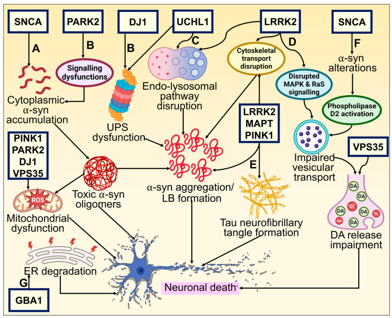
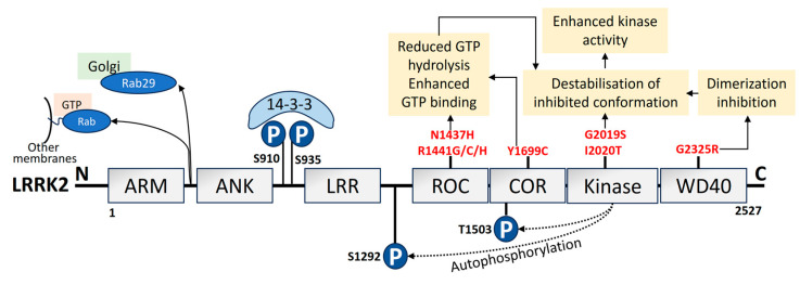
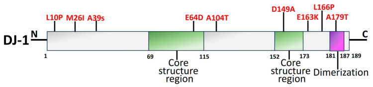
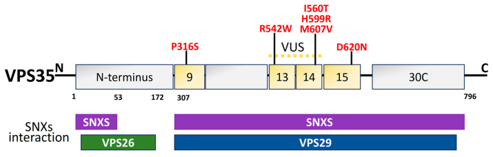
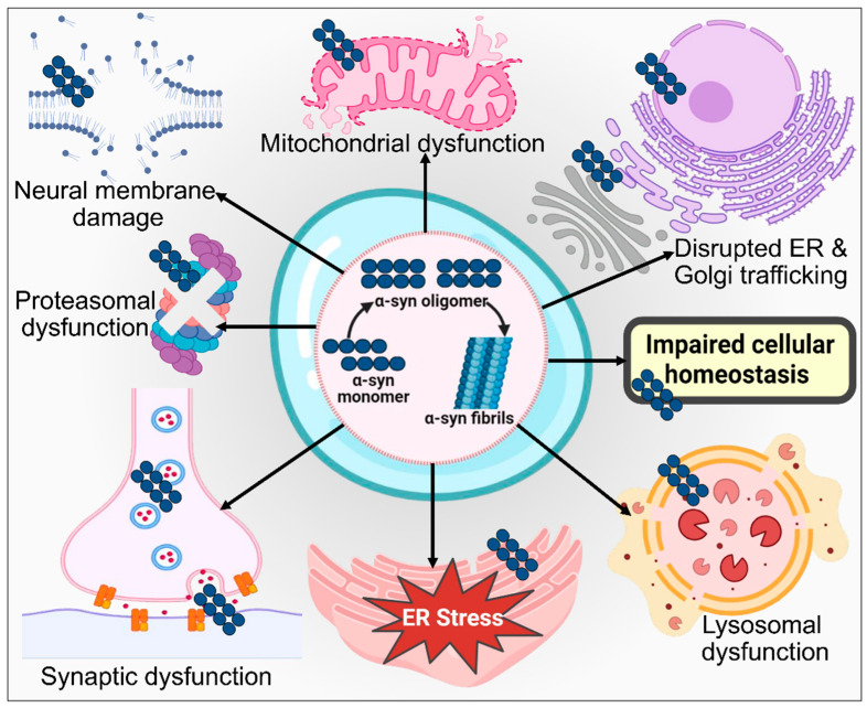
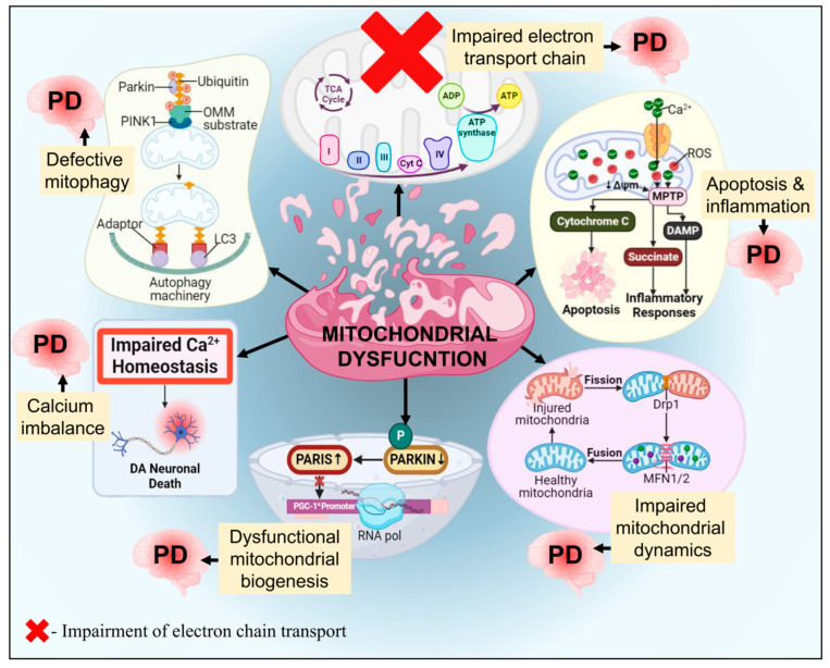
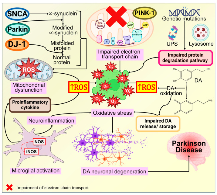
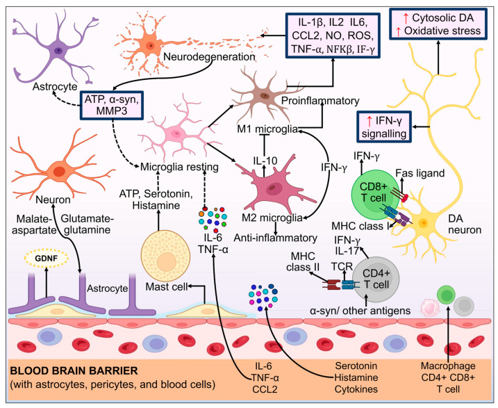
Parkinson's disease (PD) is the second most prevalent neurodegenerative movement disorder worldwide, which is primarily characterized by motor impairments. Even though multiple hypotheses have been proposed over the decades that explain the pathogenesis of PD, presently, there are no cures or promising preventive therapies for PD. This could be attributed to the intricate pathophysiology of PD and the poorly understood molecular mechanism. To address these challenges comprehensively, a thorough disease model is imperative for a nuanced understanding of PD's underlying pathogenic mechanisms. This review offers a detailed analysis of the current state of knowledge regarding the molecular mechanisms underlying the pathogenesis of PD, with a particular emphasis on the roles played by gene-based factors in the disease's development and progression. This study includes an extensive discussion of the proteins and mutations of primary genes that are linked to PD, including α-synuclein, GBA1, LRRK2, VPS35, PINK1, DJ-1, and Parkin. Further, this review explores plausible mechanisms for DAergic neural loss, non-motor and non-dopaminergic pathologies, and the risk factors associated with PD. The present study will encourage the related research fields to understand better and analyze the current status of the biochemical mechanisms of PD, which might contribute to the design and development of efficacious and safe treatment strategies for PD in future endeavors.

Keywords: Parkinson’s disease; genetics of Parkinson’s disease; molecular mechanism; risk factors; α-synuclein.


Conflict of interest statement

The authors declare no conflicts of interest.

Figures

Figure 1
Figure 1
Distinct symptoms of PD [25]. The primary symptoms of PD are mainly categorized into five types: “early symptoms, primary motor symptoms, secondary motor symptoms, primary non-motor symptoms, and secondary non-motor symptoms”.
Figure 2
Figure 2
Genetic basis of PD and its underlying molecular pathways toward neurodegeneration. (A) The neuronal defense against α-syn aggregating could consequently be compromised by mutations altering these proteins. The cytoplasmic concentration of the α-syn monomer is increased by missense mutations and chromosomal multiplications of SNCA, promoting oligomerization of α-syn that is cytotoxic. This results in neuronal membrane damage and mitochondrial dysfunction [27,94]. (B) DJ-1 and Parkin (encoded by PARK2) interact and participate in regular UPS operations [95]. Mutations affecting these proteins may reduce the neuron’s ability to respond to α-synuclein aggregation. α-synuclein aggregates that build up inside neurites and axons in late-onset PD when there is residual UPS function eventually become trapped inside a central Lewy body in surviving neurons. DJ1 also possesses antioxidative characteristics, which may offer an additional connection to α-synuclein fibrillization and impaired function [96]. (C) UCHL1 preserves a pool of monoubiquitin for E3 ligase and UPS function while inhibiting the degradation of free ubiquitin in the endosomal–lysosomal pathway [97,98]. UPS functioning and protein buildup clearance need ATP generation by mitochondria. Loss of PINK1, DJ-1, and Parkin activities significantly impairs normal mitochondrial activity, which leads to early-onset parkinsonism [99]. (D, E) Tau (encoded by microtubule-associated protein tau—MAPT) typically maintains the microtubule network in equilibrium, facilitating intracellular signaling and neuronal trafficking. Abnormal phosphorylation impairs its functionality and causes neurofibrillary tangles to develop [100,101]. Phosphorylation, intracellular signaling, and cellular trafficking all seem to be interconnected events requiring LRRK2 [95]. (F) Mutation-induced altered α-syn activity causes reduced vesicular binding, which negates the inhibition of phospholipase D2, an enzyme implicated in vesicle trafficking and lipid-mediated signaling cascades [102]. Further, restricted release of neurotransmitters and their buildup in the cytosol may produce reactive oxygen species, which in turn causes neuronal death. (G) GBA1 mutation leads to ER stress activation and degradation, which causes DAergic neuronal death [103].
Figure 3
Figure 3
Schematic depiction of α-syn structure domains. Three different domains can be distinguished from the 140-amino-acid α-syn protein. The N-terminus amphipathic domain is composed of KTKEGV repeats that contain the amino acid residues affected by the primary α-syn gene mutations (A30P, E46K, H50Q, G51D, A53T, and A53E) in PD [112,113]. Mutations linked to Parkinson’s disease (red) and phosphorylation sites (blue) have also been depicted.
Figure 4
Figure 4
Domain organization, upstream regulation, and PD-linked pathogenic mutations of LRRK2. LRRK2 is made of 2527 amino acids. It consists of 7 domains: ARM, armadillo; ANK, ankyrin; LRR, Leucine-rich repeat; ROC, Ras of complex; COR, C-terminal of ROC; kinase; and WD40 domain [168]. The phosphorylation sites in the N-terminus are pSer910 and pSer935 (blue), which mediate 14-3-3 binding to LRRK2. The autophosphorylation sites are pSer1292 and pThr1503 (blue). PD mutations (red) lead to the pathological mechanism that increases kinase activity. Upstream regulation by LRRK2, such as the pathogenic VPS35 mutation, potential LRRK2 recruitment by unidentified Rabs to other organelle membranes or vesicles, and Rab29 recruitment to the Golgi has also been depicted [169].
Figure 5
Figure 5
Schematic representation of structural domains of the mitochondrial-associated kinase PINK1, and the RBR-E3 Ubiquitin Ligase Parkin. (A) PINK1 is divided into distinct sections. Individual domains are depicted and labeled as follows: MTS, mitochondrial targeting sequence; TM, transmembrane domain; NT, N-terminal, regulatory domain; INS, insertion; CTE, C-terminal extension [185]. Depending on the residues and protein areas affected, PINK1-PD mutations (red) can be classified as having an impact on substrate binding, kinase activity, or PINK1 structure. (B) Parkin is comprised of 465 amino acids. Individual domains are depicted and labeled as follows: UBL, ubiquitin-like domain; ACT, activating element; RING, really interesting new gene domain; IBR, in-between-RING domain; REP, repressor element [186]. Mutations in PINK1 and Parkin cause PARIS accumulation that leads to neurodegeneration. Mutations linked to parkinson’s disease (red) and phosphorylation sites (blue) in PINK1 and parkin have also been depicted.
Figure 6
Figure 6
An illustration of functional domains of the DJ-1 protein with pathogenic mutations (red). The two core structural areas (green) and the dimerization region (purple) make up the DJ-1 protein. DJ-1 is a single-domain protein with 189 amino acids [208].
Figure 7
Figure 7
Schematic illustration of VPS35 structural domains and interactions along with the PD mutations (red) [208]. The dimer of sinexin (SNXs) and VPS26, VPS29, and VPS35 combine to form the retromer cargo recognition complex. For the interaction with VPS26 and VPS29, the amino acid residues 1–172 in the N-terminal region and 307–796 in the C-terminal region are significant. The N- and C-terminal regions of the amino acid residues are those that interact with the SNXs. Thirteen of the thirty-four helices projected for a structural level VPS35, a right-handed α-helix solenoid, are predicted to be in the C-terminal. In PD, several missense mutations have been discovered. The location of the VPS35 variation of unknown significance (VUS) is between exons 9 and 14. Exon 15 is the site of the pathogenic mutation (D620N).
Figure 8
Figure 8
An illustration of GBA1 structural organization along with PD mutations (red). GBA1 protein is composed of 497 amino acids, which have three primary domains: 39-residue signal peptide, the conserved catalytic domain Glyco_hydro_30 (329 amino acids), and Glyco_hydro_30C domain (30C; 62 amino acids) [233].
Figure 9
Figure 9
An illustration of the α-syn protein misfolding in the pathogenesis of PD. There are various processes by which α-syn oligomers might cause toxicity. Several protein degradation mechanisms, like autophagy and lysosomal degradation, are adversely affected by oligomers, which also actively contribute to the disruption of mitochondrial function. Additionally, oligomeric α-syn induces ER stress by manipulating the autophagy lysosomal pathways and UPS. α-syn oligomers may hinder ER-Golgi trafficking, axonal transmission, and synaptic impairment by preventing the development of SNARE complexes. Furthermore, by changing the membrane homeostasis, α-syn oligomers can directly trigger cytotoxicity. Ultimately, α-syn oligomers disrupt numerous intracellular signaling pathways and destroy organelles, which may result in neuronal death in PD.
Figure 10
Figure 10
An illustration of ER stress and UPR signaling pathways in the pathogenesis of PD. Stressful circumstances brought on by oxidative stress, infections, nutritional deficiency, and ER alterations in calcium levels can cause protein folding errors to build up in the ER. Three sensor proteins—PERK, IRE1, and ATF6—control UPR by preventing the accumulation of improperly folded proteins and enhancing ER folding ability. GRP78 binds to misfolded proteins like α-syn when the ER is stressed, causing dissociation from GRP78 activates IRE1, ATF6, and PERK. They also activate caspase-3 and force the nucleus to produce CHOP. Consequently, apoptosis occurs in PD. 1-Methyl-4-phenylpyridinium (MPP+) increases the expression of CHOP, which causes ER stress.
Figure 11
Figure 11
An illustration of DA toxicity in PD pathogenesis. Mitochondrial oxidative stress causes oxidizing DA to build up, which then causes lysosomal failure, decreased glucocerebrosidase enzyme activity, and α-syn deposits in PD neurons. Increased DA production along with impacts on the functioning of mitochondria may be simultaneously caused by higher cytosolic Ca2+ concentration through caveolin-1 (Cav1) channels.
Figure 12
Figure 12
An illustration of the potential cellular mechanism of dysfunctional mitochondria in the pathogenesis of PD. Impaired biogenesis of mitochondria, elevated generation of ROS, impaired mitophagy, compromised trafficking, malfunction of the ECT, deviations in mitochondrial dynamics, calcium imbalance, or combinational processes all contribute to mitochondrial dysfunction linked to the pathogenesis of PD. The possible complicated interaction of the numerous processes leads to a vicious cycle of escalating cellular dysfunction, which in turn causes the neurodegeneration that underpins and accelerates the pathogenesis of PD.
Figure 13
Figure 13
A systematic illustration of oxidative stress-mediated pathogenesis in PD. Several pathways and associated dysfunctions bring on rising oxidative stress because of genetic alterations in PD-related genes. Protein misfolding, mitochondrial damage, and oxidative stress are caused by mutations or modified protein expression. Free radical production and protein aggregation, particularly that of α-syn, are exacerbated by mitochondrial failure. Additionally, the chemical breakdown of DA can contribute to reactive DA quinones, which raise the amounts of ROS. Excessive oxidative stress leads to compromised UPS performance, which in turn is responsible for damaged/ misfolded protein degradation, further compromising cell viability. All of these distinct molecular mechanisms associated with oxidative stress are interlinked in the DAergic neuronal selective degeneration.
Figure 14
Figure 14
An illustration of neuroinflammation and immune dysregulation in the pathogenesis of PD. The declining strength of the immune response and the interaction between various cell types in the brain contribute to neuroinflammation. α-syn aggregation can interfere with the homeostatic functioning of neurons, astrocytes, microglia, or endothelial cells and can cause a rise in receptor response and the release of chemokines and proinflammatory cytokines. Microglial cells shift from the resting state to the activated M1 microglia, and they release proinflammatory cytokines that aid in the degeneration of DAergic neurons. Furthermore, in cross interactions with astrocytes and microglia, neuronal failure can produce α-syn, ATP, and matrix metalloproteinase-3 (MMP-3), amongst other substances, intensifying the toxic loop of neuroinflammation. Resting microglia are activated to M2 microglia by IL4 and IL13, which then downregulate M1 functionality by releasing IL10 cytokines, which have an anti-inflammatory effect on the CNS. The brain parenchyma is inhabited by CD4+ & CD8+ T cells, and these mediators or the dearth of their effective recovery mechanisms, further exacerbate the proinflammatory state.

Similar articles

Cited by

References

    1. Aarsland D., Batzu L., Halliday G.M., Geurtsen G.J., Ballard C., Ray Chaudhuri K., Weintraub D. Parkinson Disease-Associated Cognitive Impairment. Nat. Rev. Dis. Prim. 2021;7:47. doi: 10.1038/s41572-021-00280-3. - DOI - PubMed
    1. Bandres-Ciga S., Diez-Fairen M., Kim J.J., Singleton A.B. Genetics of Parkinson’s Disease: An Introspection of Its Journey towards Precision Medicine. Neurobiol. Dis. 2020;137:104782. doi: 10.1016/j.nbd.2020.104782. - DOI - PMC - PubMed
    1. Klein C., Westenberger A. Genetics of Parkinson’s Disease. Cold Spring Harb. Perspect. Med. 2012;2:a008888. doi: 10.1101/cshperspect.a008888. - DOI - PMC - PubMed
    1. Day J.O., Mullin S. The Genetics of Parkinson’s Disease and Implications for Clinical Practice. Genes. 2021;12:1006. doi: 10.3390/genes12071006. - DOI - PMC - PubMed
    1. Logroscino G., Urso D., Savica R. Descriptive Epidemiology of Neurodegenerative Diseases: What Are the Critical Questions? Neuroepidemiology. 2022;56:309–318. doi: 10.1159/000525639. - DOI - PMC - PubMed