. 2024 Jan 18;16(2):409.
doi: 10.3390/cancers16020409.

Combinational Pulsing of TAAs Enforces Dendritic Cell-Based Immunotherapy through T-Cell Proliferation and Interferon-γ Secretion in LLC1 Mouse Model

Affiliations

Combinational Pulsing of TAAs Enforces Dendritic Cell-Based Immunotherapy through T-Cell Proliferation and Interferon-γ Secretion in LLC1 Mouse Model

Jae-Ung Lee et al. Cancers (Basel). .

Abstract

NSCLC, the most common type of lung cancer, is often diagnosed late due to minimal early symptoms. Its high risk of recurrence or metastasis post-chemotherapy makes DC-based immunotherapy a promising strategy, offering targeted cancer destruction, low side effects, memory formation, and overcoming the immune evasive ability of cancers. However, the limited response to DCs pulsed with single antigens remains a significant challenge. To overcome this, we enhanced DC antigen presentation by pulsing with TAAs. Our study focused on enhancing DC-mediated immune response specificity and intensity by combinatorial pulsing of TAAs, selected for their prevalence in NSCLC. We selected four types of TAAs expressed in NSCLC and pulsed DCs with the optimal combination. Next, we administered TAAs-pulsed DCs into the LLC1 mouse model to evaluate their anti-tumor efficacy. Our results showed that TAAs-pulsed DCs significantly reduced tumor size and promoted apoptosis in tumor tissue. Moreover, TAAs-pulsed DCs significantly increased total T cells in the spleen compared to the unpulsed DCs. Additionally, in vitro stimulation of splenocytes from the TAAs-pulsed DCs showed notable T-cell proliferation and increased IFN-γ secretion. Our findings demonstrate the potential of multiple TAA pulsing to enhance the antigen-presenting capacity of DCs, thereby strengthening the immune response against tumors.

Keywords: dendritic cells; immunotherapy; non-small cell lung cancer; tumor-associated antigen.


Conflict of interest statement

The authors affiliated with Affiliation 1 and Affiliation 2 are employees of EHLBio Co., Ltd. and EHLCell Clinic, with Hong-Ki Lee serving as the CEO of both companies. They declare no competing financial interests, such as holding stocks or any other financial involvement related to the content of this paper. This study was conducted with complete scientific integrity and independence.

Figures
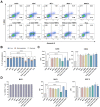
Figure 1
Figure 1
Cytotoxicity in DCs following combinational TAAs pulsing. (A) Cellular toxicity was analyzed using Annexin V/7-AAD staining. (B) The individual or combinational treatment with 2 nmol TERT increased the proportion of necrotic (7-AAD+) and early apoptotic (Annexin V+/7-AAD) cells. (C) The individual or combinational treatment with 2 nmol TERT reduced the MFI levels in CD80 and MHC I expression. (D) There were no significant changes in the MHC I-positive population following treatment with TAAs. Results are shown as mean ± SD. ** p < 0.01, *** p < 0.001, **** p < 0.0001.
Figure 2
Figure 2
Tumor response assessment following TAAs-pulsed DCs administration. (A) Experimental scheme: The LLC1 cell line was transplanted to C57BL/6 mice subcutaneously, and dendritic cells were administered into the inguinal lymph nodes. (B,C) TAAs-pulsed DCs significantly reduced the tumor volumes and weights similarly to cisplatin. (D) H&E and TUNEL analysis showed that TAAs-pulsed DCs promote tumor-specific apoptosis. These anti-cancer effects were not observed in unpulsed DCs. Results are shown as mean ± SD. * p < 0.05, ** p < 0.01, *** p < 0.001, **** p < 0.0001.
Figure 3
Figure 3
Changes in splenic T-cell subpopulations following TAAs-pulsed DCs administration. (A) Isolated splenocytes were first gated based on forward scatter (FSC) versus side scatter (SSC), and CD4+ and CD8+ subpopulations were identified from the CD3+ population. (B) LLC1 transplantation significantly increases the total number of splenocytes. (C,D) TAAs-pulsed DCs significantly increased the proportion and absolute number of total CD3+ T cells within splenocytes. (E) The proportion of CD4+ and CD8+ populations within CD3+ T cells shows no significant change. (F,G) There was a significant increase in the absolute number of CD4+ and CD8+ T cells in dendritic cells and cisplatin-administered groups, which was associated with an increase in the total number of CD3+ T cells. Results are shown as mean ± SD. * p < 0.05, ** p < 0.01, *** p < 0.001, **** p < 0.0001.
Figure 4
Figure 4
IFN-γ expression levels in splenic CD4+ and CD8+ T cells following TAAs-pulsed DCs administration. Isolated splenocytes were co-stained with IFN-γ specific fluorescent monoclonal antibody and analyzed by flow cytometry. (A,B) There were no significant differences in the proportions of CD4+ and CD8+ T cells expressing IFN-γ across all groups. (C,D) Only the TAAs-pulsed DCs significantly increased the absolute numbers of CD4+ and CD8+ T cells expressing IFN-γ compared to negative control. Results are shown as mean ± SD. * p < 0.05.
Figure 5
Figure 5
In vitro proliferation and IFN-γ secretion of isolated splenocytes after TAAs-pulsed DCs administration. Isolated splenocytes from mice were stained with CTV and further activated by PMA/Ionomycin for 3 days. Relative proliferation and IFN-γ secretion levels were analyzed using flow cytometry and ELISA. (AC) Significant activation and proliferation were observed only in the CD3+, CD4+, and CD8+ splenic T cells of mice administered with TAAs-pulsed DCs. (D) IFN-γ secretion was significantly restored in the splenocytes of mice administered with TAAs-pulsed DCs and cisplatin. Results are shown as mean ± SD. * p < 0.05, ** p < 0.01, *** p < 0.001, **** p < 0.0001.

References

    1. Ganti A.K., Klein A.B., Cotarla I., Seal B., Chou E. Update of Incidence, Prevalence, Survival, and Initial Treatment in Patients With Non-Small Cell Lung Cancer in the US. JAMA Oncol. 2021;7:1824–1832. doi: 10.1001/jamaoncol.2021.4932. - DOI - PMC - PubMed
    1. Bouleftour W., Rowinski E., Louati S., Sotton S., Wozny A.S., Moreno-Acosta P., Mery B., Rodriguez-Lafrasse C., Magne N. A Review of the Role of Hypoxia in Radioresistance in Cancer Therapy. Med. Sci. Monit. 2021;27:e934116. doi: 10.12659/MSM.934116. - DOI - PMC - PubMed
    1. Pignon J.P., Tribodet H., Scagliotti G.V., Douillard J.Y., Shepherd F.A., Stephens R.J., Dunant A., Torri V., Rosell R., Seymour L., et al. Lung adjuvant cisplatin evaluation: A pooled analysis by the LACE Collaborative Group. J. Clin. Oncol. 2008;26:3552–3559. doi: 10.1200/JCO.2007.13.9030. - DOI - PubMed
    1. Guo Q., Liu L., Chen Z., Fan Y., Zhou Y., Yuan Z., Zhang W. Current treatments for non-small cell lung cancer. Front. Oncol. 2022;12:945102. doi: 10.3389/fonc.2022.945102. - DOI - PMC - PubMed
    1. Lynch T.J., Bell D.W., Sordella R., Gurubhagavatula S., Okimoto R.A., Brannigan B.W., Harris P.L., Haserlat S.M., Supko J.G., Haluska F.G., et al. Activating mutations in the epidermal growth factor receptor underlying responsiveness of non-small-cell lung cancer to gefitinib. N. Engl. J. Med. 2004;350:2129–2139. doi: 10.1056/NEJMoa040938. - DOI - PubMed

LinkOut - more resources