. 2023 Dec 22;12(1):44.
doi: 10.3390/biomedicines12010044.

Retinal Functional Impairment in Diabetic Retinopathy

Affiliations

Retinal Functional Impairment in Diabetic Retinopathy

Cornelia Andreea Tănasie et al. Biomedicines. .

Abstract

Background: Diabetic retinopathy (DR) is a neurodegenerative disease of the retina. The aim of our study was to analyze latency changes in a full-field electroretinogram (ERG) in patients with type 2 diabetes.

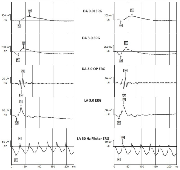
Material: This prospective study included 15 diabetic patients without DR, 16 diabetic patients with non-proliferative DR, 14 patients with pre-proliferative DR, 15 patients with proliferative DR, and 14 age-matched controls. All the participants underwent ophthalmologic examination and full-field ERGs. The ERGs were recorded with the Metrovision MonPackOne system. The latencies were analyzed for "a"- and "b"-waves in the dark-adapted (DA) 0.01 ERG, DA 3.0 ERG, DA oscillatory potentials, light-adapted (LA) 3.0 ERG, and 30 Hz flicker ERG.

Results: The delayed responses of healthy subjects compared to diabetic patients without DR were the DA oscillatory potentials (25.45 ± 1.04 ms vs. 26.15 ± 0.96 ms, p = 0.027). When comparing diabetic patients without DR and with non-proliferative DR, we did not obtain statistically significant delays. Significant delays in the DA 0.01 "b"-wave (61.91 ± 5.52 ms vs. 66.36 ± 8.12 ms, p = 0.029), DA 3.0 "b"-wave (41.01 ± 2.50 ms vs. 44.16 ± 3.78 ms, p = 0.035), and LA 3.0 "a"-wave (16.21 ± 0.91 ms vs. 16.99 ± 1.16 ms, p = 0.045) were found between non-proliferative DR and pre-proliferative DR. When comparing the groups of patients with pre-proliferative DR and proliferative DR, the LA 3.0 ERG "b"-wave (32. 63 ± 2.53 ms vs. 36.19 ± 3.21 ms, p < 0.0001), LA 30 Hz flicker ERG "a"-wave (19.56 ± 3.59 vs. 21.75 ± 4.74 ms, p= 0.025), and "b"-wave (32.23 ± 4.02 vs. 36.68 ± 3.48 ms, p = 0.017) were delayed.

Conclusions: the electrophysiological findings from our study indicate that there is a substantial dysfunction of the neural retina in all stages of DR.

Keywords: diabetes mellitus; diabetic retinopathy; full-field ERG.


Conflict of interest statement

The authors declare no conflict of interest.

Figures

Figure 1
Figure 1
Standard ERG in a healthy individual. DA—dark adapted; LA—light adapted; RE—right eye; LE—left eye; A1—a-wave; B1—b-wave; O1—OP N1; O2—OP N2.
Figure 2
Figure 2
Comparison of mean values of l DA 0.01 a-wave latencies between normal subjects (control) and the subgroups of patients with diabetes mellitus without DR (DR-), patients with non-proliferative diabetic retinopathy (NDR), patients with pre-proliferative diabetic retinopathy (PPDR), and patients with proliferative diabetic retinopathy (PDR). * p < 0.05; ** p < 0.01.
Figure 3
Figure 3
Comparison of mean values of DA 0.01 b-wave latencies between normal subjects (control) and the groups of patients with diabetes mellitus without DR (DR-), patients with non-proliferative diabetic retinopathy (NDR), patients with pre-proliferative diabetic retinopathy (PPDR), and patients with proliferative diabetic retinopathy (PDR). * p < 0.05; ** p < 0.01; *** p < 0.001.
Figure 4
Figure 4
Comparison of mean values of DA 3.0 a-wave latencies between healthy subjects (control) and the subgroups of patients with diabetes mellitus without DR (DR-), patients with non-proliferative diabetic retinopathy (NDR), patients with pre-proliferative diabetic retinopathy (PPDR), and patients with proliferative diabetic retinopathy (PDR).
Figure 5
Figure 5
Comparison of mean values of DA 3.0 b-wave latencies between healthy subjects (control) and the groups of patients with diabetes mellitus without DR (DR-), patients with non-proliferative diabetic retinopathy (NDR), patients with pre-proliferative diabetic retinopathy (PPDR), and patients with proliferative diabetic retinopathy (PDR). * p < 0.05; ** p < 0.01; *** p < 0.001.
Figure 6
Figure 6
Comparison of mean values of DA 3.0 OP N1 wave latencies between healthy subjects (control) and the groups of patients with diabetes mellitus without DR (DR-), patients with non-proliferative diabetic retinopathy (NDR), patients with pre-proliferative diabetic retinopathy (PPDR), and patients with proliferative diabetic retinopathy (PDR). * p < 0.05; ** p < 0.01; *** p < 0.001.
Figure 7
Figure 7
Comparison of mean values of DA 3.0 OP N2 wave latencies between healthy subjects (control) and the groups of patients with diabetes mellitus without DR (DR-), patients with non-proliferative diabetic retinopathy (NDR), patients with pre-proliferative diabetic retinopathy (PPDR), and patients with proliferative diabetic retinopathy (PDR). * p < 0.05; ** p < 0.01; *** p < 0.001.
Figure 8
Figure 8
Comparison of mean values of LA 3.0 a-wave latencies between healthy subjects (control) and the groups of patients with diabetes mellitus without DR (DR-), patients with non-proliferative diabetic retinopathy (NDR), patients with pre-proliferative diabetic retinopathy (PPDR), and patients with proliferative diabetic retinopathy (PDR). * p < 0.05; ** p < 0.01.
Figure 9
Figure 9
Comparison of mean values of LA 3.0 b-wave latencies between healthy subjects (control) and the groups of patients with diabetes mellitus without DR (DR-), patients with non-proliferative diabetic retinopathy (NDR), patients with pre-proliferative diabetic retinopathy (PPDR), and patients with proliferative diabetic retinopathy (PDR). *** p < 0.001.
Figure 10
Figure 10
Comparison of mean values of LA 30 Hz flicker a-wave latencies between healthy subjects (control) and the groups of patients with diabetes mellitus without DR (DR-), patients with non-proliferative diabetic retinopathy (NDR), patients with pre-proliferative diabetic retinopathy (PPDR), and patients with proliferative diabetic retinopathy (PDR). * p < 0.05; ** p < 0.01.
Figure 11
Figure 11
Comparison of mean values of LA 30 Hz flicker b-wave latencies between healthy subjects (control) and the groups of patients with diabetes mellitus without DR (DR-), patients with non-proliferative diabetic retinopathy (NDR), patients with pre-proliferative diabetic retinopathy (PPDR), and patients with proliferative diabetic retinopathy (PDR). * p < 0.05; ** p < 0.01; *** p < 0.001.

References

    1. Flaxman S.R., Bourne R.R.A., Resnikoff S., Ackland P., Braithwaite T., Cicinelli M.V., Das A., Jonas J.B., Keeffe J., Kempen J.H., et al. Global causes of blindness and distance vision impairment 1990–2020: A systematic review and meta-analysis. Lancet Glob. Health. 2017;5:1221–1234. doi: 10.1016/S2214-109X(17)30393-5. - DOI - PubMed
    1. Engelgau M.M., Geiss L.S., Saaddine J.B., Boyle J.P., Benjamin S.M., Gregg E.W., Tierney E.F., Rios-Burrows N., Mokdad A.H., Ford E.S., et al. The Evolving diabetes burden in the United States. Ann. Intern. Med. 2004;140:945–950. doi: 10.7326/0003-4819-140-11-200406010-00035. - DOI - PubMed
    1. Tyrberg M., Lindblad U., Melander A., Lövestam-Adrian M., Ponjavic V., Andréasson S. Electrophysiological studies in newly onset type 2 diabetes without visible vascular retinopathy. Doc. Ophthalmol. 2011;123:193–198. doi: 10.1007/s10633-011-9298-6. - DOI - PubMed
    1. Tonadea D., Kern T.S. Photoreceptor cells and RPE contribute to the development of diabetic retinopathy. Prog. Retin. Eye Res. 2021;83:1009–1019. doi: 10.1016/j.preteyeres.2020.100919. - DOI - PMC - PubMed
    1. Barber A.J., Antonetti D.A., Kern T.S., Reiter C.E.N., Soans R.S., Krady J.K., Levison S.W., Gardner T.W., Bronson S.K. The Ins2Akita mouse as a model of early retinal com-plications in diabetes. Investig. Ophthalmol. Vis. Sci. 2005;46:2210–2218. doi: 10.1167/iovs.04-1340. - DOI - PubMed

LinkOut - more resources