Review
. 2024 Jan 8;12(1):124.
doi: 10.3390/biomedicines12010124.

Involvement of Lysophospholipids in Pulmonary Vascular Functions and Diseases

Affiliations
Review

Involvement of Lysophospholipids in Pulmonary Vascular Functions and Diseases

Hiroaki Kume et al. Biomedicines. .

Abstract

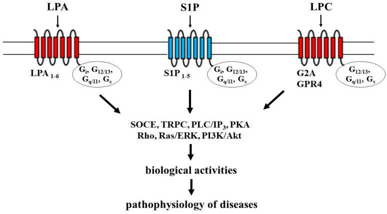
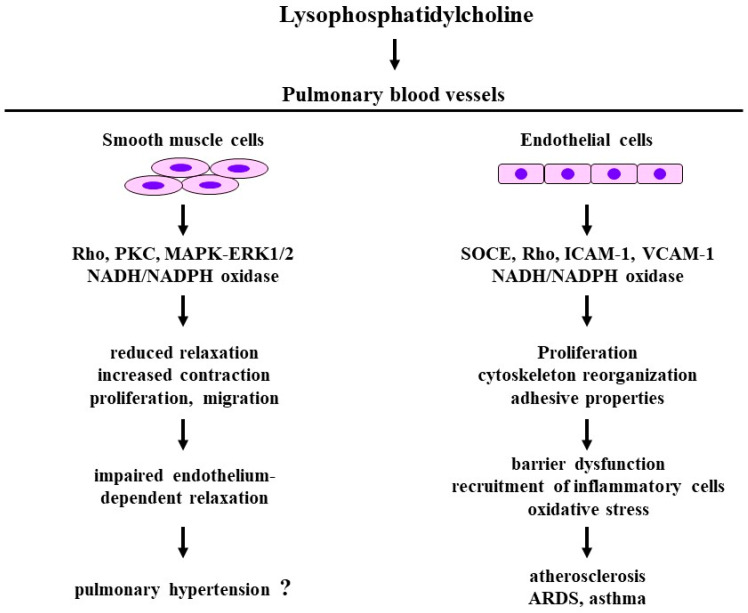
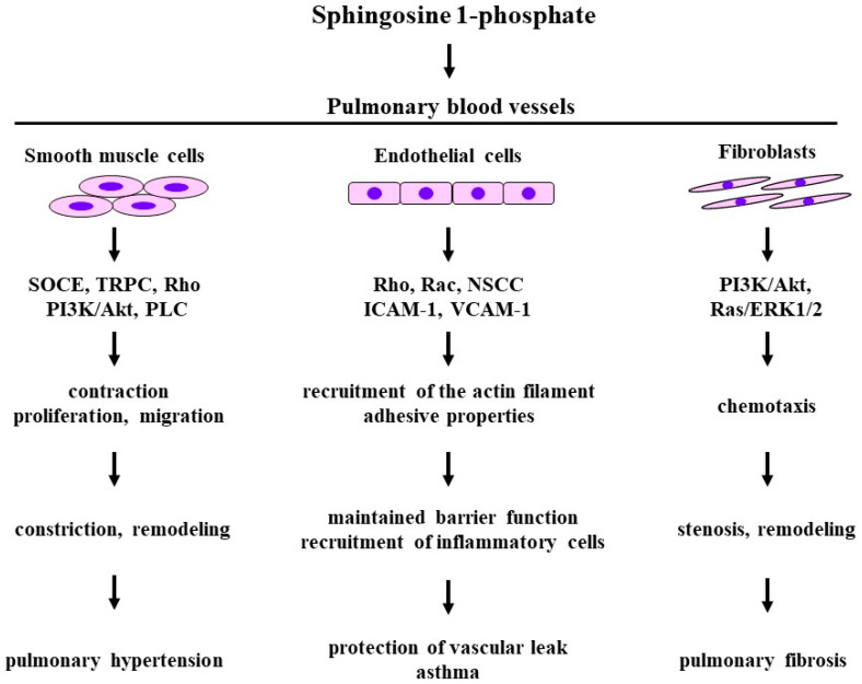
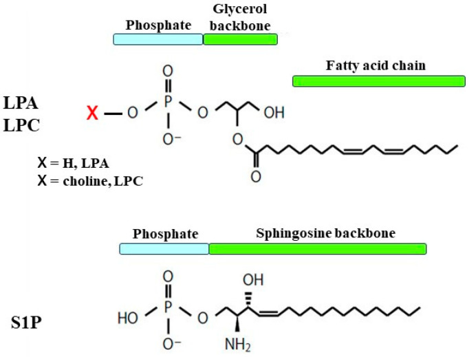
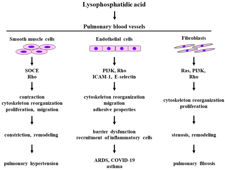
Extracellular lysophospholipids (lysophosphatidic acid, lysophosphatidylcholine, sphingosine 1-phosphate, etc.), which are synthesized from phospholipids in the cell membrane, act as lipid mediators, and mediate various cellular responses in constituent cells in the respiratory system, such as contraction, proliferation, migration, and cytoskeletal organization. In addition to these effects, the expression of the adhesion molecules is enhanced by these extracellular lysophospholipids in pulmonary endothelial cells. These effects are exerted via specific G protein-coupled receptors. Rho, Ras, and phospholipase C (PLC) have been proven to be their signaling pathways, related to Ca2+ signaling due to Ca2+ dynamics and Ca2+ sensitization. Therefore, lysophospholipids probably induce pulmonary vascular remodeling through phenotype changes in smooth muscle cells, endothelial cells, and fibroblasts, likely resulting in acute respiratory distress syndrome due to vascular leak, pulmonary hypertension, and pulmonary fibrosis. Moreover, lysophospholipids induce the recruitment of inflammatory cells to the lungs via the enhancement of adhesion molecules in endothelial cells, potentially leading to the development of asthma. These results demonstrate that lysophospholipids may be novel therapeutic targets not only for injury, fibrosis, and hypertension in the lung, but also for asthma. In this review, we discuss the mechanisms of the effects of lysophospholipids on the respiratory system, and the possibility of precision medicine targeting lysophospholipids as treatable traits of these diseases.

Keywords: ARDS; asthma; lysophosphadidylcholine; lysophosphatidic acid; pulmonary endothelial cells; pulmonary fibroblasts; pulmonary fibrosis; pulmonary hypertension; pulmonary vascular smooth muscle; sphingosine 1-phosphate.


Conflict of interest statement

The authors declare no conflict of interest.

Figures

Figure 2
Figure 2
The receptors and intracellular signal transduction processes of lysophospholipids. LPA: lysophosphatidic acid; LPC: lysophosphatidylcholine; S1P: sphingosine 1-phosphate; SOCE: store-operated calcium entry; TRPC: transient receptor potential channels; PLC: phospholipase C; IP3: inositol 1,4,5-trisphosphate; PKA: protein kinase A; ERK: extracellular signal-regulated kinase; PI3K: phosphoinositol 3-kinase; Akt: protein kinase B. Illustrated based on Refs. [20,21,22,24,25].
Figure 4
Figure 4
The effects of lysophosphatidylcholine on the constituent cells and related molecular mechanisms in pulmonary blood vessels. Disease arising from pathophysiology based on physiological activities induced by lysophosphatidic acid. SOCE: store-operated calcium entry; PKC: protein kinase C; MAPK: mitogen-activated protein kinase; ERK: extracellular signal-regulated kinase; PI3K: phosphoinositol 3-kinase; ICAM-1: intercellular adhesion molecule 1; VCAM-1: vascular cell adhesion molecule-1. The 1st row: the constituent cells; the 2nd row: molecular mechanisms; the 3rd row: biological activities; the 4th row: pathophysiology; the 5th row: diseases related to pulmonary vasculatures. Illustrated based on Refs. [4,16,18,52,53,54,57,58,59,60,61,62,64,65,66,67,68,69,70,71,75,76,77,78,79,80,81,82,83,86,87,88,89,90].
Figure 5
Figure 5
The effects of sphingosine 1-phosphate on the constituent cells and related molecular mechanisms in pulmonary blood vessels. Disease arising from pathophysiology based on physiological activities induced by lysophosphatidic acid. SOCE: store-operated calcium entry; TRPC: transient receptor potential channels; NSCC: non-selective cation channels; ERK: extracellular signal-regulated kinase; PI3K: phosphoinositol 3-kinase; PLC: phospholipase C; ICAM-1: intercellular adhesion molecule 1; VCAM-1: vascular cell adhesion molecule-1. The 1st row: the constituent cells; the 2nd row: molecular mechanisms; the 3rd row: biological activities; the 4th row: pathophysiology; the 5th row: diseases related to pulmonary vasculatures. Illustrated based on Refs. [10,13,96,97,101,102,104,105,106,107,108,109,110,111,112,113,114,115,116,117,118,122,123,124,125,127,128,129,130,131,132,133,134,135,136,137,138,139].
Figure 1
Figure 1
The representative structures of lysophospholipids. LPA: lysophosphatidic acid; LPC: lysophosphatidylcholine; S1P: sphingosine 1-phosphate. The number of carbons and double bonds varies depending on each fatty acid.
Figure 3
Figure 3
The effects of lysophosphatidic acid on the constituent cells and related molecular mechanisms in pulmonary blood vessels. Disease arising from pathophysiology based on physiological activities induced by lysophosphatidic acid. SOCE: store-operated calcium entry; PI3K: phosphoinositol 3-kinase; ICAM-1: intercellular adhesion molecule 1. The 1st row: the constituent cells; the 2nd row: molecular mechanisms; the 3rd row: biological activities; the 4th row: pathophysiology; the 5th row: diseases related to pulmonary vasculature. Illustrated based on Refs. [5,9,11,14,15,17,24,29,30,32,33,34,35,36,37,38,39,40,41,42,43,44,45,46,47,48].

Similar articles

Cited by

References

    1. Rivera R., Chun J. Biological effects of lysophospholipids. Rev. Physiol. Biochem. Pharmacol. 2008;160:25–46. doi: 10.1007/112_0507. - DOI - PubMed
    1. Poli G., Leonarduzzi G., Biasi F., Chiarpotto E. Oxidative stress and cell signalling. Curr. Med. Chem. 2004;11:1163–1182. doi: 10.2174/0929867043365323. - DOI - PubMed
    1. Ueda N. A Rheostat of Ceramide and Sphingosine-1-Phosphate as a Determinant of Oxidative Stress-Mediated Kidney Injury. Int. J. Mol Sci. 2022;23:4010. doi: 10.3390/ijms23074010. - DOI - PMC - PubMed
    1. Liu P., Zhu W., Chen C., Yan B., Zhu L., Chen X., Peng C. The mechanisms of lysophosphatidylcholine in the development of diseases. Life Sci. 2020;247:117443. doi: 10.1016/j.lfs.2020.117443. - DOI - PubMed
    1. Nikitopoulou I., Fanidis D., Ntatsoulis K., Moulos P., Mpekoulis G., Evangelidou M., Vassiliou A.G., Dimakopoulou V., Jahaj E., Tsipilis S., et al. Increased Autotaxin Levels in Severe COVID-19, Correlating with IL-6 Levels, Endothelial Dysfunction Biomarkers, and Impaired Functions of Dendritic Cells. Int. J. Mol. Sci. 2021;22:10006. doi: 10.3390/ijms221810006. - DOI - PMC - PubMed

LinkOut - more resources