

Platelet-Rich Plasma in Dermatology: New Insights on the Cellular Mechanism of Skin Repair and Regeneration
- PMID: 38255655
- PMCID: PMC10817627
- DOI: 10.3390/life14010040
Platelet-Rich Plasma in Dermatology: New Insights on the Cellular Mechanism of Skin Repair and Regeneration
Abstract
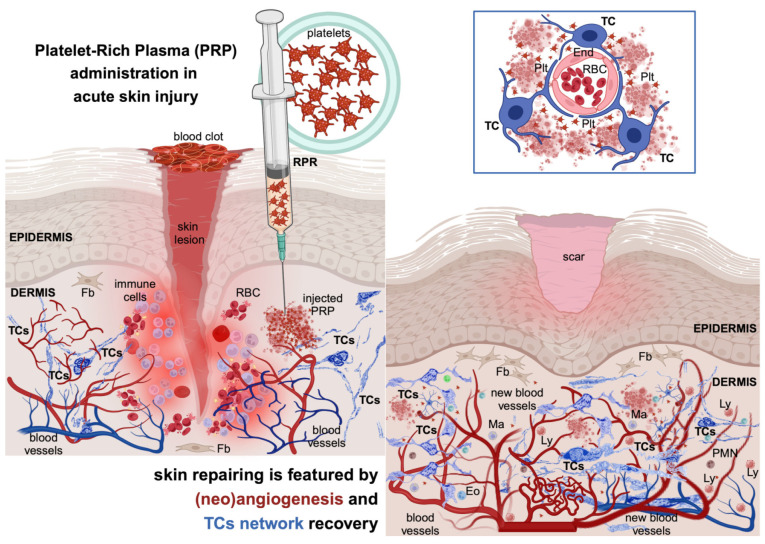
The skin's recognised functions may undergo physiological alterations due to ageing, manifesting as varying degrees of facial wrinkles, diminished tautness, density, and volume. Additionally, these functions can be disrupted (patho)physiologically through various physical and chemical injuries, including surgical trauma, accidents, or chronic conditions like ulcers associated with diabetes mellitus, venous insufficiency, or obesity. Advancements in therapeutic interventions that boost the skin's innate regenerative abilities could significantly enhance patient care protocols. The application of Platelet-Rich Plasma (PRP) is widely recognized for its aesthetic and functional benefits to the skin. Yet, the endorsement of PRP's advantages often borders on the dogmatic, with its efficacy commonly ascribed solely to the activation of fibroblasts by the factors contained within platelet granules. PRP therapy is a cornerstone of regenerative medicine which involves the autologous delivery of conditioned plasma enriched by platelets. This is achieved by centrifugation, removing erythrocytes while retaining platelets and their granules. Despite its widespread use, the precise sequences of cellular activation, the specific cellular players, and the molecular machinery that drive PRP-facilitated healing are still enigmatic. There is still a paucity of definitive and robust studies elucidating these mechanisms. In recent years, telocytes (TCs)-a unique dermal cell population-have shown promising potential for tissue regeneration in various organs, including the dermis. TCs' participation in neo-angiogenesis, akin to that attributed to PRP, and their role in tissue remodelling and repair processes within the interstitia of several organs (including the dermis), offer intriguing insights. Their potential to contribute to, or possibly orchestrate, the skin regeneration process following PRP treatment has elicited considerable interest. Therefore, pursuing a comprehensive understanding of the cellular and molecular mechanisms at work, particularly those involving TCs, their temporal involvement in structural recovery following injury, and the interconnected biological events in skin wound healing and regeneration represents a compelling field of study.
Keywords: aesthetic dermatology; autologous transplant; platelet-rich plasma; skin regeneration; skin repairing; telocytes.
Conflict of interest statement
The authors declare no conflict of interest.
Figures
Similar articles
-
Skin Telocytes Could Fundament the Cellular Mechanisms of Wound Healing in Platelet-Rich Plasma Administration.Cells. 2024 Aug 8;13(16):1321. doi: 10.3390/cells13161321. Cells. 2024. PMID: 39195210 Free PMC article. Review.
-
Profound Properties of Protein-Rich, Platelet-Rich Plasma Matrices as Novel, Multi-Purpose Biological Platforms in Tissue Repair, Regeneration, and Wound Healing.Int J Mol Sci. 2024 Jul 19;25(14):7914. doi: 10.3390/ijms25147914. Int J Mol Sci. 2024. PMID: 39063156 Free PMC article. Review.
-
The Use of Platelet-Rich Plasma in Aesthetic and Regenerative Medicine: A Comprehensive Review.Aesthetic Plast Surg. 2019 Jun;43(3):803-814. doi: 10.1007/s00266-018-1293-9. Epub 2018 Dec 14. Aesthetic Plast Surg. 2019. PMID: 30552470 Review.
-
Platelets in wound healing and regenerative medicine.Platelets. 2018 Sep;29(6):556-568. doi: 10.1080/09537104.2018.1430357. Epub 2018 Feb 14. Platelets. 2018. PMID: 29442539 Review.
-
Dermal Telocytes: A Different Viewpoint of Skin Repairing and Regeneration.Cells. 2022 Dec 2;11(23):3903. doi: 10.3390/cells11233903. Cells. 2022. PMID: 36497161 Free PMC article. Review.
Cited by
-
Efficacy of mesenchymal stem cells and platelet-rich plasma therapies on wound healing: A Systematic Review and meta-analysis.Regen Ther. 2025 May 19;30:75-91. doi: 10.1016/j.reth.2025.04.010. eCollection 2025 Dec. Regen Ther. 2025. PMID: 40491558 Free PMC article. Review.
-
The use of platelet rich plasma in the treatment of degenerative joint disease in cats: an exploratory case series.Front Vet Sci. 2024 May 28;11:1394055. doi: 10.3389/fvets.2024.1394055. eCollection 2024. Front Vet Sci. 2024. PMID: 38863451 Free PMC article.
-
Platelet-Rich Concentrates in the Management of Lichen Planus-A Comprehensive Review.J Clin Med. 2025 Jul 29;14(15):5368. doi: 10.3390/jcm14155368. J Clin Med. 2025. PMID: 40806989 Free PMC article. Review.
-
Skin Telocytes Could Fundament the Cellular Mechanisms of Wound Healing in Platelet-Rich Plasma Administration.Cells. 2024 Aug 8;13(16):1321. doi: 10.3390/cells13161321. Cells. 2024. PMID: 39195210 Free PMC article. Review.
-
Aesthetic Rehabilitation Medicine: Enhancing Wellbeing beyond Functional Recovery.Medicina (Kaunas). 2024 Apr 5;60(4):603. doi: 10.3390/medicina60040603. Medicina (Kaunas). 2024. PMID: 38674249 Free PMC article. Review.
References
Publication types
LinkOut - more resources
Full Text Sources
Medical
Research Materials
