iPSC-Derived Endothelial Cells Reveal LDLR Dysfunction and Dysregulated Gene Expression Profiles in Familial Hypercholesterolemia
- PMID: 38255763
- PMCID: PMC10815294
- DOI: 10.3390/ijms25020689
iPSC-Derived Endothelial Cells Reveal LDLR Dysfunction and Dysregulated Gene Expression Profiles in Familial Hypercholesterolemia
Abstract
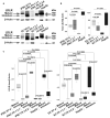
Defects in the low-density lipoprotein receptor (LDLR) are associated with familial hypercholesterolemia (FH), manifested by atherosclerosis and cardiovascular disease. LDLR deficiency in hepatocytes leads to elevated blood cholesterol levels, which damage vascular cells, especially endothelial cells, through oxidative stress and inflammation. However, the distinctions between endothelial cells from individuals with normal and defective LDLR are not yet fully understood. In this study, we obtained and examined endothelial derivatives of induced pluripotent stem cells (iPSCs) generated previously from conditionally healthy donors and compound heterozygous FH patients carrying pathogenic LDLR alleles. In normal iPSC-derived endothelial cells (iPSC-ECs), we detected the LDLR protein predominantly in its mature form, whereas iPSC-ECs from FH patients have reduced levels of mature LDLR and show abolished low-density lipoprotein uptake. RNA-seq of mutant LDLR iPSC-ECs revealed a unique transcriptome profile with downregulated genes related to monocarboxylic acid transport, exocytosis, and cell adhesion, whereas upregulated signaling pathways were involved in cell secretion and leukocyte activation. Overall, these findings suggest that LDLR defects increase the susceptibility of endothelial cells to inflammation and oxidative stress. In combination with elevated extrinsic cholesterol levels, this may result in accelerated endothelial dysfunction, contributing to early progression of atherosclerosis and other cardiovascular pathologies associated with FH.
Keywords: LDLR; directed differentiation; endothelial cells; familial hypercholesterolemia; patient-specific iPSCs.
Conflict of interest statement
The authors declare no conflicts of interest.
Figures


Similar articles
-
Low-density lipoprotein receptor-deficient hepatocytes differentiated from induced pluripotent stem cells allow familial hypercholesterolemia modeling, CRISPR/Cas-mediated genetic correction, and productive hepatitis C virus infection.Stem Cell Res Ther. 2019 Jul 29;10(1):221. doi: 10.1186/s13287-019-1342-6. Stem Cell Res Ther. 2019. PMID: 31358055 Free PMC article.
-
JD induced pluripotent stem cell-derived hepatocytes faithfully recapitulate the pathophysiology of familial hypercholesterolemia.Hepatology. 2012 Dec;56(6):2163-71. doi: 10.1002/hep.25871. Epub 2012 Sep 17. Hepatology. 2012. PMID: 22653811 Free PMC article.
-
Induced pluripotent stem cell line ICGi037-A, obtained by reprogramming peripheral blood mononuclear cells from a patient with familial hypercholesterolemia due to heterozygous p.Trp443Arg mutations in LDLR.Stem Cell Res. 2022 Apr;60:102703. doi: 10.1016/j.scr.2022.102703. Epub 2022 Feb 6. Stem Cell Res. 2022. PMID: 35152179
-
The molecular genetic basis and diagnosis of familial hypercholesterolemia in Denmark.Dan Med Bull. 2002 Nov;49(4):318-45. Dan Med Bull. 2002. PMID: 12553167 Review.
-
Homozygous familial hypercholesterolaemia: update on management.Paediatr Int Child Health. 2016 Nov;36(4):243-247. doi: 10.1080/20469047.2016.1246640. Paediatr Int Child Health. 2016. PMID: 27967828 Review.
Cited by
-
Bridging the Gap: Endothelial Dysfunction and the Role of iPSC-Derived Endothelial Cells in Disease Modeling.Int J Mol Sci. 2024 Dec 11;25(24):13275. doi: 10.3390/ijms252413275. Int J Mol Sci. 2024. PMID: 39769040 Free PMC article. Review.
References
MeSH terms
Substances
Grants and funding
LinkOut - more resources
Full Text Sources
Medical
Miscellaneous

