. 2024 Jan 5;25(2):728.
doi: 10.3390/ijms25020728.

Evaluation of the LDN-0060609 PERK Inhibitor as a Selective Treatment for Primary Open-Angle Glaucoma: An In Vitro Study on Human Retinal Astrocytes

Affiliations

Evaluation of the LDN-0060609 PERK Inhibitor as a Selective Treatment for Primary Open-Angle Glaucoma: An In Vitro Study on Human Retinal Astrocytes

Wioletta Rozpędek-Kamińska et al. Int J Mol Sci. .

Abstract

The term glaucoma encompasses various neurodegenerative eye disorders, among which the most common is primary open-angle glaucoma (POAG). Recently, the essential role of human retinal astrocytes (HRA) in glaucoma progression has been placed in the spotlight. It has been found that placing the endoplasmic reticulum (ER) under stress and activating PERK leads to apoptosis of HRA cells, which inhibits their neuroprotective effect in the course of glaucoma. Therefore, the aim of the present study was to evaluate the effectiveness of the small-molecule PERK inhibitor LDN-0060609 in countering ER stress conditions induced in HRA cells in vitro. The activity of LDN-0060609 was studied in terms of protein and mRNA expression, cytotoxicity, genotoxicity, caspase-3 level and cell cycle progression. LDN-0060609 at 25 μM proved to be a potent inhibitor of the major PERK substrate, p-eIF2α (49% inhibition). The compound markedly decreased the expression of pro-apoptotic ER stress-related genes (ATF4, DDIT3, BAX and Bcl-2). Treatment with LDN-0060609 significantly increased cell viability, decreased genotoxicity and caspase-3 levels, and restored cell cycle distribution in HRA cells with activated ER stress conditions. These findings indicate that the small-molecule PERK inhibitor LDN-0060609 can potentially be developed into a novel anti-glaucoma agent.

Keywords: PERK; PERK inhibitor; eIF2α; endoplasmic reticulum stress; glaucoma; glaucoma treatment; unfolded protein response.


Conflict of interest statement

The authors declare no conflicts of interest.

Figures

Figure 1
Figure 1
Western blot analysis (A) and quantification measured by means of optical densitometry (B) of eIF2α phosphorylation in HRA cells treated with the LDN-0060609 PERK inhibitor and Th (thapsigargin) as an ER stress inducer. All experiments were performed in triplicate, values are expressed as mean ± SEM, n = 3. *** p < 0.001 versus the positive control (Th). C—negative control (untreated HRA cells); Th—thapsigargin-treated HRA cells (ER-stressed HRA).
Figure 2
Figure 2
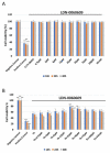
Evaluation of the mRNA expression of the ER stress-related apoptotic genes: (A) ATF4, (B) DDIT3, (C) BAX, (D) Bcl-2 in HRA cells treated with the small-molecule PERK inhibitor LDN-0060609 alone, Th alone, or with both Th and LDN-0060609. The analysis was performed using a TaqMan gene expression assay. All experiments were performed in triplicate, and values are expressed as mean ± SEM, n = 3. *** p < 0.001 versus Th. Control—untreated HRA cells; 0.1% DMSO—HRA cells treated with solvent 0.1% dimethyl sulfoxide; Th—thapsigargin-treated HRA cells (ER-stressed HRA).
Figure 2
Figure 2
Evaluation of the mRNA expression of the ER stress-related apoptotic genes: (A) ATF4, (B) DDIT3, (C) BAX, (D) Bcl-2 in HRA cells treated with the small-molecule PERK inhibitor LDN-0060609 alone, Th alone, or with both Th and LDN-0060609. The analysis was performed using a TaqMan gene expression assay. All experiments were performed in triplicate, and values are expressed as mean ± SEM, n = 3. *** p < 0.001 versus Th. Control—untreated HRA cells; 0.1% DMSO—HRA cells treated with solvent 0.1% dimethyl sulfoxide; Th—thapsigargin-treated HRA cells (ER-stressed HRA).
Figure 3
Figure 3
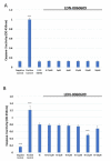
Analysis of the cytotoxicity of the LDN-0060609 compound toward HRA cells (A) and viability of ER-stressed HRA cells upon treatment with LDN-0060609 PERK inhibitor (B), as assessed by the colorimetric XTT assay. All experiments were performed in triplicate; values are expressed as mean ± SEM, n = 3. ** p < 0.01; *** p < 0.001 versus negative control (A) and Th (B). Negative control—untreated HRA cells; positive control—HRA cells treated with 99.9% dimethyl sulfoxide; 0.1% DMSO—HRA cells treated with the solvent, 0.1% dimethyl sulfoxide; Th—thapsigargin-treated HRA cells (ER-stressed HRA).
Figure 4
Figure 4
Genotoxicity analysis in HRA cells treated with the LDN-0060609 PERK inhibitor (A) and in ER-stressed HRA cells treated with LDN-0060609 (B), as indicated by the comet assay. All experiments were performed in triplicate. The value of cells scored for individual experiments was 100. Box plots show the median, first and third quartiles, minimum and maximum values. ** p < 0.01; *** p < 0.001 versus the negative control (A) and versus Th (B). Negative control—untreated HRA cells; Positive control—HRA cells treated with 99.9% dimethyl sulfoxide; 0.1% DMSO—HRA cells treated with the solvent, 0.1% dimethyl sulfoxide; Th—thapsigargin-treated HRA cells (ER-stressed HRA).
Figure 5
Figure 5
Assessment of the level of apoptosis in HRA cells (A) or ER-stressed HRA cells (B) following exposure to LDN-0060609. Results were determined by a caspase-3 assay. All experiments were performed in triplicate; values are expressed as mean ± SEM, n = 3. *** p < 0.001 versus the negative control (A) and versus Th (B). Negative control—untreated HRA cells; Positive control—HRA cells treated with 1 μM staurosporine; 0.1% DMSO—HRA cells treated with the solvent, 0.1% dimethyl sulfoxide; Th—thapsigargin-treated HRA cells (ER-stressed HRA).
Figure 6
Figure 6
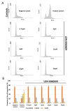
Flow cytometric analysis with propidium iodide (PI) staining of cell cycle progression in HRA cells exposed to LDN-0060609 alone (A,B) and ER-stressed HRA cells exposed to LDN-0060609 (C,D). The results are presented as histograms of DNA content (A,C) and bar graphs showing cell cycle distribution determined from the DNA content histograms (B,D). All experiments were performed in triplicate; values are expressed as mean ± SEM, n = 3. * p < 0.05, ** p < 0.01, *** p < 0.001 versus the negative control (B) and versus Th (D). Negative control—untreated HRA cells; Positive control—HRA cells treated with 1 μM nocodazole; Th—thapsigargin-treated HRA cells (ER-stressed HRA).
Figure 6
Figure 6
Flow cytometric analysis with propidium iodide (PI) staining of cell cycle progression in HRA cells exposed to LDN-0060609 alone (A,B) and ER-stressed HRA cells exposed to LDN-0060609 (C,D). The results are presented as histograms of DNA content (A,C) and bar graphs showing cell cycle distribution determined from the DNA content histograms (B,D). All experiments were performed in triplicate; values are expressed as mean ± SEM, n = 3. * p < 0.05, ** p < 0.01, *** p < 0.001 versus the negative control (B) and versus Th (D). Negative control—untreated HRA cells; Positive control—HRA cells treated with 1 μM nocodazole; Th—thapsigargin-treated HRA cells (ER-stressed HRA).

Similar articles

Cited by

References

    1. Jayaram H., Kolko M., Friedman D.S., Gazzard G. Glaucoma: Now and Beyond. Lancet. 2023;402:1788–1801. doi: 10.1016/S0140-6736(23)01289-8. - DOI - PubMed
    1. Tham Y.C., Li X., Wong T.Y., Quigley H.A., Aung T., Cheng C.Y. Global Prevalence of Glaucoma and Projections of Glaucoma Burden through 2040: A Systematic Review and Meta-Analysis. Ophthalmology. 2014;121:2081–2090. doi: 10.1016/j.ophtha.2014.05.013. - DOI - PubMed
    1. Burton M.J., Ramke J., Marques A.P., Bourne R.R.A., Congdon N., Jones I., Ah Tong B.A.M., Arunga S., Bachani D., Bascaran C., et al. The Lancet Global Health Commission on Global Eye Health: Vision beyond 2020. Lancet Glob. Health. 2021;9:e489–e551. doi: 10.1016/S2214-109X(20)30488-5. - DOI - PMC - PubMed
    1. McLaughlin T., Medina A., Perkins J., Yera M., Wang J.J., Zhang S.X. Cellular Stress Signaling and the Unfolded Protein Response in Retinal Degeneration: Mechanisms and Therapeutic Implications. Mol. Neurodegener. 2022;17:25. doi: 10.1186/s13024-022-00528-w. - DOI - PMC - PubMed
    1. Chen X., Shi C., He M., Xiong S., Xia X. Endoplasmic Reticulum Stress: Molecular Mechanism and Therapeutic Targets. Signal Transduct. Target. Ther. 2023;8:352. doi: 10.1038/s41392-023-01570-w. - DOI - PMC - PubMed