. 2024 Jan 6;25(2):754.
doi: 10.3390/ijms25020754.

First Evidence of Mineralocorticoid Receptor Gene and Protein Expression in Rat and Human Thyroid Tissues and Cell Cultures

Affiliations

First Evidence of Mineralocorticoid Receptor Gene and Protein Expression in Rat and Human Thyroid Tissues and Cell Cultures

Jacopo Manso et al. Int J Mol Sci. .

Abstract

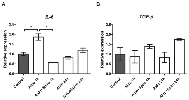
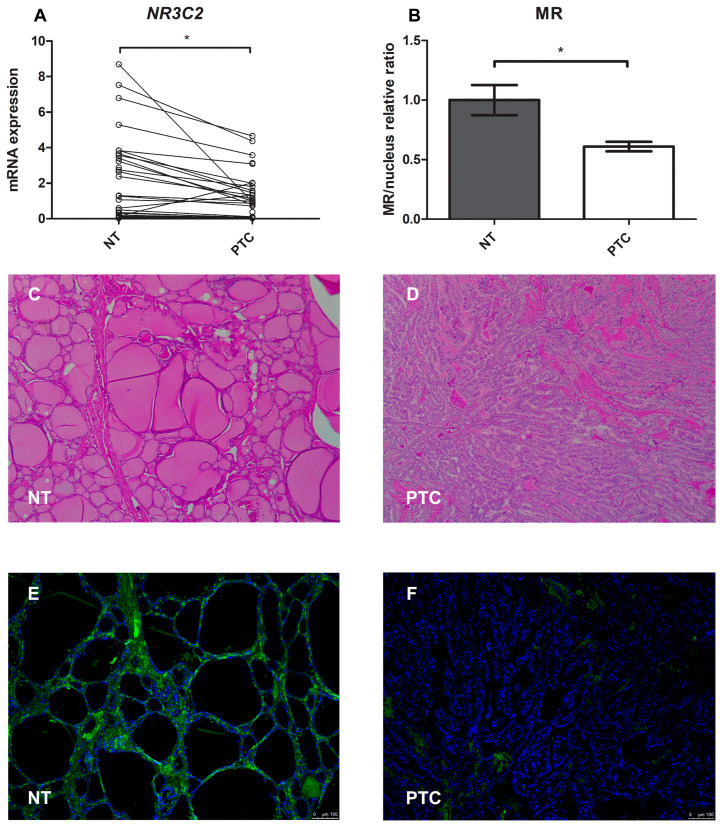
Aldosterone (Aldo) exerts its action through binding with the mineralocorticoid receptor (MR). Clinically, a link between primary aldosteronism (PA) and thyroid diseases has been hypothesised. However, the presence and activity of MR on the thyroid have not yet been demonstrated. We investigated the gene/protein expression and activation of MR in primary thyroid cell cultures (normal rat thyroid [FRTL-5] and human papillary thyroid cancer [PTC] cell lines, BCPAP and K1) through qRT-PCR analysis, immunofluorescence, and confocal microscopy. We also studied the effects of Aldo on thyroid-specific and inflammation genes in vitro. Paired human normal and neoplastic thyroid tissues were also studied. We demonstrated both gene and protein expression and activation of MR in normal rat thyroid and human PTC lines. Incubation with Aldo induced an acute increase in IL-6 expression in both the FRTL-5 and BCPAP lines, which was antagonised by spironolactone, and an acute and late upregulation of thyroid-specific genes in FRTL-5. MR was also expressed at both gene and protein levels in normal human thyroid tissues and in PTC, with a progressive decline during neoplastic tumourigenesis, particularly in more aggressive histotypes. We present the first evidence of MR gene and protein expression in both normal and pathological thyroid cells and tissues. We have shown that MR is present and functionally activated in thyroid tissue. Binding of Aldo to MR induces the expression of inflammatory and thyroid-specific genes, and the thyroid may thus be considered a novel mineralocorticoid target tissue.

Keywords: aldosterone; mineralocorticoid; mineralocorticoid receptor; papillary thyroid cancer; thyroid; thyroid cancer.


Conflict of interest statement

The authors declare that the research was conducted in the absence of any commercial or financial relationships that could be construed as a potential conflict of interest.

Figures

Figure 1
Figure 1
(A,B) NR3C2 mRNA expression levels assessed by quantitative real-time polymerase chain reaction. (A) NR3C2 expression analysis in different rat tissue types; (B) NR3C2 mRNA expression levels in different cell lines. Experiments were performed in triplicate. ** = p < 0.01, * = p < 0.05.
Figure 2
Figure 2
Confocal microscopy showing MR protein expression, localisation, and activation in the FRTL-5 cell line. Nuclear translocation of MR in FRTL-5 treated with aldosterone (1 µM for 24 h) can be seen in the expanded image detail. MR is shown in green; DNA stained with DRAQ5™ is shown in blue. Z-interval: 1 μm. Magnification: 63× oil immersion objective. Scale bar = 25 μm. MR = mineralocorticoid receptor.
Figure 3
Figure 3
(A,B) mRNA expression levels in the FRTL-5 cell line assessed by quantitative real-time polymerase chain reaction. (A) IL-6 gene expression; (B) TGF-β gene expression. Aldo 1 h = Aldosterone 1 µM for 1 h; Aldo+Spiro 1 h = Aldosterone 1 µM combined with Spironolactone 10 µM for 1 h; Aldo 24 h = Aldosterone 1 µM for 24 h; Aldo+Spiro 24 h = Aldosterone 1 µM combined with Spironolactone 10 µM for 24 h. Experiments were performed in triplicate. * = p < 0.001.
Figure 4
Figure 4
(A–C) mRNA expression levels in the FRTL-5 cell line assessed by quantitative real-time polymerase chain reaction. (A) Thyroid peroxidase gene expression levels; (B) Thyroglobulin gene expression levels; (C) SLC5A5 (NIS) gene expression levels. Aldo 1 h = Aldosterone 1 µM for 1 h; Aldo+Spiro 1 h = Aldosterone 1 µM combined with Spironolactone 10 µM for 1 h; Aldo 24 h = Aldosterone 1 µM for 24 h; Aldo+Spiro 24 h = Aldosterone 1 µM combined with Spironolactone 10 µM for 24 h; TPO = thyroid peroxidase; TG = thyroglobulin. Experiments were performed in triplicate. * = p < 0.01.
Figure 5
Figure 5
Confocal microscopy showing MR protein expression, localisation, and activation in BCPAP. Nuclear translocation of MR in BCPAP treated with aldosterone can be seen in the expanded image detail. MR is shown in green; DNA stained with DRAQ5™ is shown in blue. Z-interval 1 μm. Magnification: 63× oil immersion objective. Scale bar = 25 μm. MR = mineralocorticoid receptor.
Figure 6
Figure 6
Confocal microscopy showing MR protein expression, localisation, and activation in K1. The mineralocorticoid receptor is shown in green; DNA stained with DRAQ5™ is shown in blue. Z-interval: 1 μm. Magnification: 63× oil immersion objective. Scale bar = 25 μm. MR = mineralocorticoid receptor.
Figure 7
Figure 7
(A,B) IL-6 mRNA expression levels in the PTC (BCPAP and K1) cell lines assessed by quantitative real-time polymerase chain reaction. (A) IL-6 gene expression in the BCPAP cell line; (B) IL-6 gene expression in the K1 cell line. Aldo 1 h = Aldosterone 1 µM for 1 h; Aldo+Spiro 1 h = Aldosterone 1 µM combined with Spironolactone 10 µM for 1 h; Aldo 24 h = Aldosterone 1 µM for 24 h; Aldo+Spiro 24 h = Aldosterone 1 µM combined with Spironolactone 10 µM for 24 h. Experiments were performed in triplicate. * = p < 0.001.
Figure 8
Figure 8
(AF) Mineralocorticoid receptor gene and protein expression in normal human thyroid tissue compared with papillary thyroid cancer tissue. (A) Representation of the Wilcoxon test for paired data on NR3C2 mRNA expression: each PTC tissue sample was compared with its healthy tissue counterpart; (B) Quantification of mean fluorescence intensity (MFI); (C,D) Representative images of the immunohistochemistry analysis of normal thyroid tissue and papillary thyroid cancer tissue stained with hematoxylin and eosin; (E,F) Immunofluorescence of normal thyroid tissue and papillary thyroid cancer tissue (green = MR, blue = nucleus). Magnification: objective 10×. Scale bar = 100 μm. NT = normal thyroid tissue; PTC = papillary thyroid cancer. * = p < 0.001.
Figure 9
Figure 9
Western blot analysis for 11β-Hydroxysteroid dehydrogenase type 2. (A) Representative Western blot analysis for different tissue samples. (B) Quantification of band intensity, Samples 1–3: healthy counterparts of human kidney carcinoma specimens; Samples 4, 5: healthy counterparts of human papillary thyroid cancer. 11βHSD2 = 11β-Hydroxysteroid dehydrogenase type 2.
Figure 10
Figure 10
NR3C2 mRNA expression levels in different human thyroid cancer tissues assessed by quantitative real-time polymerase chain reaction. PTC = classic variant of papillary thyroid cancer; ATC = anaplastic thyroid cancer. * = p < 0.001.

References

    1. Balsamo A., Cicognani A., Gennari M., Sippell W.G., Menabo S., Baronio F., Riepe F.G. Functional characterization of naturally occurring NR3C2 gene mutations in Italian patients suffering from pseudohypoaldosteronism type 1. Eur. J. Endocrinol. 2007;156:249–256. doi: 10.1530/eje.1.02330. - DOI - PubMed
    1. Booth R.E., Johnson J.P., Stockand J.D. Aldosterone. Adv. Physiol. Educ. 2002;26:8–20. doi: 10.1152/advan.00051.2001. - DOI - PubMed
    1. Patel P.D., Sherman T.G., Goldman D.J., Watson S.J. Molecular cloning of a mineralocorticoid (type I) receptor complementary DNA from rat hippocampus. Mol. Endocrinol. 1989;3:1877–1885. doi: 10.1210/mend-3-11-1877. - DOI - PubMed
    1. Pearce P., Funder J.W. High affinity aldosterone binding sites (type I receptors) in rat heart. Clin. Exp. Pharmacol. Physiol. 1987;14:859–866. doi: 10.1111/j.1440-1681.1987.tb02422.x. - DOI - PubMed
    1. Funder J.W., Pearce P.T., Smith R., Smith A.I. Mineralocorticoid action: Target tissue specificity is enzyme, not receptor, mediated. Science. 1988;242:583–585. doi: 10.1126/science.2845584. - DOI - PubMed

LinkOut - more resources