. 2024 Jan 10;25(2):845.
doi: 10.3390/ijms25020845.

Effect of a New Substance with Pyridoindole Structure on Adult Neurogenesis, Shape of Neurons, and Behavioral Outcomes in a Chronic Mild Stress Model in Rats

Affiliations

Effect of a New Substance with Pyridoindole Structure on Adult Neurogenesis, Shape of Neurons, and Behavioral Outcomes in a Chronic Mild Stress Model in Rats

Alexandra Zvozilova et al. Int J Mol Sci. .

Abstract

Despite an accumulating number of studies, treatments for depression are currently insufficient. Therefore, the search for new substances with antidepressant potential is very important. In this study, we hypothesized that treatment with a newly synthesized pyridoindole derivative compound SMe1EC2M3 would result in protective and antidepressant-like effects on behavioral outcomes and reverse the impaired adult hippocampal neurogenesis caused by chronic mild stress (CMS). We found that chronic administration of 5 mg/kg and 25 mg/kg SMe1EC2M3 to adult Sprague Dawley rats ameliorated the consequences of CMS on immobility and swimming time in a forced swim test. A slight sedative effect of the highest dose of SMe1EC2M3 in the nonstress group was observed in the open field. SMe1EC2M3 in the highest dose ameliorated CMS-induced decreases in the sucrose preference test. Administration of SMe1EC2M3 significantly increased SOX2-positive cells in the hippocampal dentate gyrus (DG) in CMS compared to control animals. A significant reduction in glial fibrillary acid protein (GFAP)-positive cells in the DG of CMS compared to control animals was observed. Administration of both 5 and 25 mg/kg SMe1EC2M3 significantly increased signal of GFAP-positive cells in the DG of CMS animals. No such effects of SMe1EC2M3 were observed in the cornu ammonis hippocampal area. Additionally, we found that incubation of primary hippocampal neurons in the presence of 1.50 µM SMe1EC2M3 significantly stimulated the length of neurites. Overall, we found that the negative effects of CMS on depression-like behavior are partially reduced by the administration of SMe1EC2M3 and are associated with changes in hippocampal neurogenesis and neuronal differentiation. SMe1EC2M3 represents a potential drug candidate with positive neuroplastic effects and neurogenesis-associated effects in therapeutic approaches to depression.

Keywords: animal model; antidepressants; behavior; immunohistochemistry; major depressive disorder; pyridoindoles; triple reuptake inhibitors.


Conflict of interest statement

The authors declare no conflict of interest.

Figures

Figure 1
Figure 1
Structural formula of (±)-cis ethyl 8-methoxy-6-methyl-3,4,4a,5,9bH-hexahydro-1Hpyrido[4,3-b]indole-2-carboxylate (SMe1EC2M3).
Figure 2
Figure 2
The effect of SMe1EC2M3 on sucrose preference. Means are represented in box plots ± SEM (n = 10–12 animals/group). Two-way ANOVA revealed main effect of stress vs. nonstress conditions (p ≤ 0.05) and main effect of treatment (p ≤ 0.01). Fisher’s LSD post hoc test revealed significantly different values, marked with * p ≤ 0.05, compared to nonstress vehicle group, and with # p ≤ 0.05 compared to stress vehicle group.
Figure 3
Figure 3
The effect of SMe1EC2M3 on distance traveled in open field test. Means are represented in box plots ± SEM (n = 11–12 animals/group). Two-way ANOVA revealed main effect of stress vs. nonstress conditions (p ≤ 0.01) and an interaction of treatment and conditions (p ≤ 0.05). Fisher’s LSD post hoc test revealed significantly different values, marked with * p ≤ 0.05; ** p ≤ 0.01 compared to nonstress vehicle group.
Figure 4
Figure 4
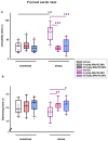
The effect of SMe1EC2M3 on immobility (a) and swimming time (b) in forced swim test. Means are represented in box plots ± SEM (n = 10–12 animals/group). Two-way ANOVA revealed main effect of treatment (p ≤ 0.001), stress vs. nonstress conditions (p ≤ 0.01), and their interactions (p ≤ 0.001) on immobility time (a) and main effect of treatment (p ≤ 0.05) on swimming time (b). Fisher’s LSD post hoc test revealed significantly different values, marked with *** p ≤ 0.001 compared to nonstress vehicle group; marked with # p ≤ 0.05; ## p ≤ 0.01; ### p ≤ 0.001 compared to stress vehicle group.
Figure 5
Figure 5
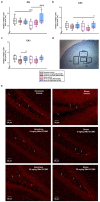
SMe1EC2M3 restores the number of neural progenitor cells (SOX2+) in the hippocampal areas DG (a), CA3 (b), and CA1 (c) in Sprague Dawley rats in a chronic mild stress animal model of depression. Representative image of chosen regions (d) and representative microscopic images (20× magnification) showing immunoreactivity to SOX2 (red; white arrows) in the DG (e). SOX2+ cells were detected in three different regions of interest (ROIs; 1 × 105 µm2) in the DG, CA3, and CA1 parts of the hippocampus (n = 6 animals/group, 8 sections of bilateral hippocampus/group, 3 ROIs/each part of the hippocampus). Means are represented in box plots ± SEM. Two-way ANOVA revealed main effect of the factors interaction (p ≤ 0.001) and treatment (p ≤ 0.001) for the DG region (a) and main effect of treatment for the CA1 and CA3 regions (p ≤ 0.01). Tukey–Kramer post hoc revealed significantly different values, marked with * p ≤ 0.05; *** p ≤ 0.001 compared to nonstress vehicle group; ### p ≤ 0.001 compared to stress vehicle group.
Figure 6
Figure 6
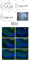
SMe1EC2M3 restores the number of glial cells (GFAP+) in the hippocampal areas DG (a), CA3 (b), and CA1 (c) in Sprague Dawley rats in a chronic mild stress animal model of depression. Representative image of chosen regions (d) and representative microscopic images (20× magnification) showing immunoreactivity to GFAP (green; white arrows) and DAPI (blue) in the DG (e). GFAP+ cells were detected in three different regions of interest (ROIs; 1 × 105 µm2) in the DG, CA3, and CA1 parts of the hippocampus (n = 6 animals/group, 8 sections of bilateral hippocampus/group, 3 ROIs/each part of the hippocampus). Means are represented in box plots ± SEM. Two-way ANOVA revealed main effect of the factors interaction (p ≤ 0.001), stress vs. nonstress condition (p ≤ 0.05), and treatment (p ≤ 0.001) for the DG region. Tukey–Kramer post hoc revealed significantly different values marked with * p ≤ 0.05; *** p ≤ 0.001 compared to nonstress vehicle group; # p ≤ 0.05 compared to stress vehicle group.
Figure 7
Figure 7
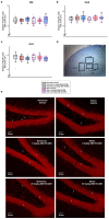
Effect of SMe1EC2M3 on the number of neurons (NeuN+) in the hippocampal areas DG (a), CA3 (b), and CA1 (c) in Sprague Dawley rats in a chronic mild stress animal model of depression. Representative image of chosen regions (d) and representative microscopic images (20× magnification) showing immunoreactivity to NeuN (red; white arrows) in the DG (e). NeuN+ cells were detected in three different regions of interest (ROIs; 1 × 105 µm2) in the DG, CA3, and CA1 parts of the hippocampus (n = 6 animals/group, 8 sections of bilateral hippocampus/group, 3 ROIs/each part of the hippocampus). Means are represented in box plots ± SEM.
Figure 8
Figure 8
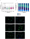
Quantitative assessment of neurite outgrowth in primary hippocampal neurons isolated from P0 Wistar rats in response to different concentrations of SMe1EC2M3 and 10 µM ATRA on DIV9. Neurite length was quantified in all neurons in the visual field from the edge of the nucleus to the apical end of the neurite. Cells were plated at a density of 0.8 × 10⁵/mL (n = 6 pups). Six coverslips per experimental group (n = 100–150 cells) and at least seven areas of interest per coverslip were evaluated. The average value of the longest neurite is shown. Means are represented in box plots ± SEM. ANOVA revealed significant differences between groups. Tukey–Kramer post hoc revealed significantly different values marked with * p ≤ 0.05; ** p ≤ 0.01 compared to untreated CTRL group (a). Stacked bar graphs showing the percentage of neurons with a defined length of neurites (b). Representative fluorescent microscopic images of neurons in experimental groups (c). Cells labeled MAP2 (green) and nucleus (blue). DIV = day in vitro.
Figure 9
Figure 9
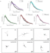
Sholl analysis of neurite arborization in primary hippocampal neurons isolated from P0 Wistar rats in response to different concentrations of SMe1EC2M3 and 10 µM ATRA on DIV9. The number of neurite intersections for concentric circles with various distances far from the nucleus was evaluated using FIJI/IMAGE J. Cells were plated at a density of 0.8 × 10⁵/mL (n = 6 pups) (a). Representative binary images of neurons for all experimental groups are shown (n = 40 cells per group) (b). Scale bar represents 50 µm. DIV = day in vitro.
Figure 10
Figure 10
Experimental design. Experimental schedule of chronic mild stress applied for three weeks, antidepressant treatment with SMe1EC2M3 in two doses for two consecutive weeks, and behavioral testing schedule. CMS—chronic mild stress, FST—forced swim test, OF—open field test, SPT—sucrose preference test.

References

    1. Andersen I., Thielen K., Bech P., Nygaard E., Diderichsen F. Increasing prevalence of depression from 2000 to 2006. Scand. J. Public Health. 2011;39:857–863. doi: 10.1177/1403494811424611. - DOI - PubMed
    1. Delgado P.L. Depression: The case for a monoamine deficiency. J. Clin. Psychiatry. 2000;61((Suppl. S6)):7–11. - PubMed
    1. Thase M.E., Entsuah A.R., Rudolph R.L. Remission rates during treatment with venlafaxine or selective serotonin reuptake inhibitors. Br. J. Psychiatry. 2001;178:234–241. doi: 10.1192/bjp.178.3.234. - DOI - PubMed
    1. Kennedy S.H., Young A.H., Blier P. Strategies to achieve clinical effectiveness: Refining existing therapies and pursuing emerging targets. J. Affect. Disord. 2011;132((Suppl. S1)):S21–S28. doi: 10.1016/j.jad.2011.03.048. - DOI - PubMed
    1. Perez-Caballero L., Torres-Sanchez S., Romero-López-Alberca C., González-Saiz F., Mico J.A., Berrocoso E. Monoaminergic system and depression. Cell Tissue Res. 2019;377:107–113. doi: 10.1007/s00441-018-2978-8. - DOI - PubMed

Substances

LinkOut - more resources