

Autophagy Markers Are Altered in Alzheimer's Disease, Dementia with Lewy Bodies and Frontotemporal Dementia
- PMID: 38256197
- PMCID: PMC10816165
- DOI: 10.3390/ijms25021125
Autophagy Markers Are Altered in Alzheimer's Disease, Dementia with Lewy Bodies and Frontotemporal Dementia
Abstract
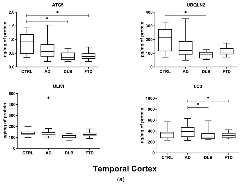
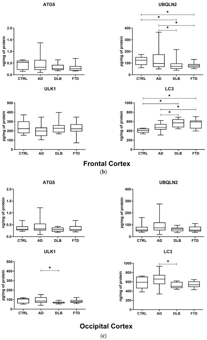
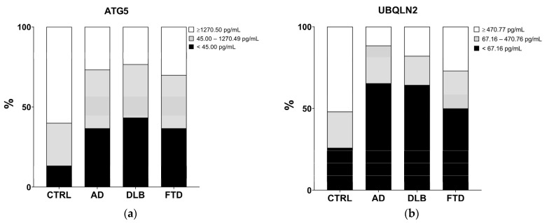
The accumulation of protein aggregates defines distinct, yet overlapping pathologies such as Alzheimer's disease (AD), dementia with Lewy bodies (DLB), and frontotemporal dementia (FTD). In this study, we investigated ATG5, UBQLN2, ULK1, and LC3 concentrations in 66 brain specimens and 120 plasma samples from AD, DLB, FTD, and control subjects (CTRL). Protein concentration was measured with ELISA kits in temporal, frontal, and occipital cortex specimens of 32 AD, 10 DLB, 10 FTD, and 14 CTRL, and in plasma samples of 30 AD, 30 DLB, 30 FTD, and 30 CTRL. We found alterations in ATG5, UBQLN2, ULK1, and LC3 levels in patients; ATG5 and UBQLN2 levels were decreased in both brain specimens and plasma samples of patients compared to those of the CTRL, while LC3 levels were increased in the frontal cortex of DLB and FTD patients. In this study, we demonstrate alterations in different steps related to ATG5, UBQLN2, and LC3 autophagy pathways in DLB and FTD patients. Molecular alterations in the autophagic processes could play a role in a shared pathway involved in the pathogenesis of neurodegeneration, supporting the hypothesis of a common molecular mechanism underlying major neurodegenerative dementias and suggesting different potential therapeutic targets in the autophagy pathway for these disorders.
Keywords: ATG5; Alzheimer’s disease; LC3; UBQLN2; ULK1; autophagy; brain; dementia with Lewy bodies; frontotemporal dementia; neurodegeneration.
Conflict of interest statement
The authors declare no conflicts of interest. The funders had no role in the design of the study; in the collection, analyses, or interpretation of data; in the writing of the manuscript, or in the decision to publish the results.
Figures
Similar articles
-
Comparisons of clinical symptoms in biomarker-confirmed Alzheimer's disease, dementia with Lewy bodies, and frontotemporal dementia patients in a local memory clinic.Psychogeriatrics. 2015 Dec;15(4):235-41. doi: 10.1111/psyg.12103. Epub 2014 Dec 23. Psychogeriatrics. 2015. PMID: 25533477
-
Relative Incidence of Seizures and Myoclonus in Alzheimer's Disease, Dementia with Lewy Bodies, and Frontotemporal Dementia.J Alzheimers Dis. 2017;60(1):211-223. doi: 10.3233/JAD-170031. J Alzheimers Dis. 2017. PMID: 28826176 Free PMC article.
-
Distinct perfusion patterns in Alzheimer's disease, frontotemporal dementia and dementia with Lewy bodies.Eur Radiol. 2014 Sep;24(9):2326-33. doi: 10.1007/s00330-014-3172-3. Epub 2014 Jul 5. Eur Radiol. 2014. PMID: 24996793
-
Autophagy and Alzheimer's Disease.Adv Exp Med Biol. 2020;1207:3-19. doi: 10.1007/978-981-15-4272-5_1. Adv Exp Med Biol. 2020. PMID: 32671736 Review.
-
Molecular and Anatomical Imaging of Dementia With Lewy Bodies and Frontotemporal Lobar Degeneration.Semin Nucl Med. 2021 May;51(3):264-274. doi: 10.1053/j.semnuclmed.2020.12.002. Epub 2021 Jan 2. Semin Nucl Med. 2021. PMID: 33402272 Review.
Cited by
-
KIF9 Ameliorates Neuropathology and Cognitive Dysfunction by Promoting Macroautophagy in a Mouse Model of Alzheimer's Disease.Aging Cell. 2025 May;24(5):e14490. doi: 10.1111/acel.14490. Epub 2025 Jan 19. Aging Cell. 2025. PMID: 39829171 Free PMC article.
-
Current strategies in the management of dementia with lewy bodies and future directions based on disease pathophysiology.Mol Neurodegener. 2025 Jul 1;20(1):78. doi: 10.1186/s13024-025-00856-7. Mol Neurodegener. 2025. PMID: 40598239 Free PMC article. Review.
-
Dysregulated microRNAs in blood correlate with central nervous system neuropathology of prion disease.Vet Res. 2025 Jul 1;56(1):132. doi: 10.1186/s13567-025-01566-0. Vet Res. 2025. PMID: 40597437 Free PMC article.
-
Synergistic anticancer effects of camptothecin and sotorasib in KRAS-mutated pancreatic ductal adenocarcinoma.Front Pharmacol. 2025 Jul 18;16:1635449. doi: 10.3389/fphar.2025.1635449. eCollection 2025. Front Pharmacol. 2025. PMID: 40756996 Free PMC article.
-
The role of autophagy in the pathogenesis and treatment of multiple sclerosis.Autophagy Rep. 2025 Jul 22;4(1):2529196. doi: 10.1080/27694127.2025.2529196. eCollection 2025. Autophagy Rep. 2025. PMID: 40717814 Free PMC article. Review.
References
-
- Zhong X., Hou L., Luo X., Shi H., Hu G., He H., Chen X., Zheng D., Zhang Y., Tan Y., et al. Alterations of CSF Cystatin C Levels and their Correlations with CSF Aβ40 and Aβ42 Levels in Patients with Alzheimer’s Disease, Dementia with Lewy Bodies and the Atrophic Form of General Paresis. PLoS ONE. 2013;8:e55328. doi: 10.1371/journal.pone.0055328. - DOI - PMC - PubMed
