

Dopamine Signaling in Substantia Nigra and Its Impact on Locomotor Function-Not a New Concept, but Neglected Reality
- PMID: 38256204
- PMCID: PMC10815979
- DOI: 10.3390/ijms25021131
Dopamine Signaling in Substantia Nigra and Its Impact on Locomotor Function-Not a New Concept, but Neglected Reality
Abstract
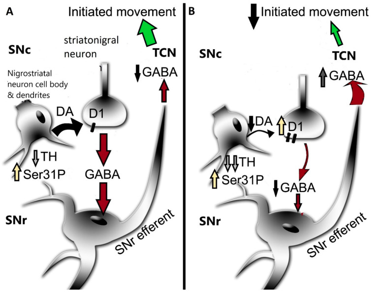
The mechanistic influences of dopamine (DA) signaling and impact on motor function are nearly always interpreted from changes in nigrostriatal neuron terminals in striatum. This is a standard practice in studies of human Parkinson's disease (PD) and aging and related animal models of PD and aging-related parkinsonism. However, despite dozens of studies indicating an ambiguous relationship between changes in striatal DA signaling and motor phenotype, this perseverating focus on striatum continues. Although DA release in substantia nigra (SN) was first reported almost 50 years ago, assessment of nigral DA signaling changes in relation to motor function is rarely considered. Whereas DA signaling has been well-characterized in striatum at all five steps of neurotransmission (biosynthesis and turnover, storage, release, reuptake, and post-synaptic binding) in the nigrostriatal pathway, the depth of such interrogations in the SN, outside of cell counts, is sparse. However, there is sufficient evidence that these steps in DA neurotransmission in the SN are operational and regulated autonomously from striatum and are present in human PD and aging and related animal models. To complete our understanding of how nigrostriatal DA signaling affects motor function, it is past time to include interrogation of nigral DA signaling. This brief review highlights evidence that changes in nigral DA signaling at each step in DA neurotransmission are autonomous from those in striatum and changes in the SN alone can influence locomotor function. Accordingly, for full characterization of how nigrostriatal DA signaling affects locomotor activity, interrogation of DA signaling in SN is essential.
Keywords: Parkinson’s disease; aging; dopamine; dopamine receptor; nigrostriatal; phosphorylation; reuptake; striatum; substantia nigra; tyrosine hydroxylase.
Conflict of interest statement
The author declares no conflict of interest.
Figures


Similar articles
-
ser31 Tyrosine hydroxylase phosphorylation parallels differences in dopamine recovery in nigrostriatal pathway following 6-OHDA lesion.J Neurochem. 2014 May;129(3):548-58. doi: 10.1111/jnc.12652. Epub 2014 Jan 27. J Neurochem. 2014. PMID: 24410633 Free PMC article.
-
Modulation of nigral dopamine signaling mitigates parkinsonian signs of aging: evidence from intervention with calorie restriction or inhibition of dopamine uptake.Geroscience. 2023 Feb;45(1):45-63. doi: 10.1007/s11357-022-00583-7. Epub 2022 May 30. Geroscience. 2023. PMID: 35635679 Free PMC article.
-
Differential expression of RET and GDNF family receptor, GFR-α1, between striatum and substantia nigra following nigrostriatal lesion: A case for diminished GDNF-signaling.Exp Neurol. 2023 Aug;366:114435. doi: 10.1016/j.expneurol.2023.114435. Epub 2023 May 12. Exp Neurol. 2023. PMID: 37178997
-
Converging roles of ion channels, calcium, metabolic stress, and activity pattern of Substantia nigra dopaminergic neurons in health and Parkinson's disease.J Neurochem. 2016 Oct;139 Suppl 1(Suppl Suppl 1):156-178. doi: 10.1111/jnc.13572. Epub 2016 Mar 23. J Neurochem. 2016. PMID: 26865375 Free PMC article. Review.
-
Toward full restoration of synaptic and terminal function of the dopaminergic system in Parkinson's disease by stem cells.Ann Neurol. 2003;53 Suppl 3:S135-46; discussion S146-8. doi: 10.1002/ana.10482. Ann Neurol. 2003. PMID: 12666105 Review.
Cited by
-
Placebo effects of repetitive transcranial magnetic stimulation on negative symptoms and cognition in patients with schizophrenia spectrum disorders: a systematic review and meta-analysis.Front Psychiatry. 2024 May 28;15:1377257. doi: 10.3389/fpsyt.2024.1377257. eCollection 2024. Front Psychiatry. 2024. PMID: 38863608 Free PMC article.
-
Gray matter structural and functional brain abnormalities in Parkinson's disease: a meta-analysis of VBM and ALFF data.J Neurol. 2025 Mar 19;272(4):276. doi: 10.1007/s00415-025-12934-3. J Neurol. 2025. PMID: 40106017 Review.
-
Olfactory Projections to Locomotor Control Centers in the Sea Lamprey.Int J Mol Sci. 2024 Aug 29;25(17):9370. doi: 10.3390/ijms25179370. Int J Mol Sci. 2024. PMID: 39273317 Free PMC article.
-
Glial Derived Neurotrophic Factor and Schizophrenia Spectrum Disorders: A Scoping Review.Curr Neuropharmacol. 2025;23(5):564-578. doi: 10.2174/011570159X340124241205095729. Curr Neuropharmacol. 2025. PMID: 39679463 Free PMC article.
References
-
- Glowinski J., Axelrod J., Iversen L.L. Regional studies of catecholamines in the rat brain. IV. Effects of drugs on the disposition and metabolism of H3-norepinephrine and H3-dopamine. J. Pharmacol. Exp. Ther. 1966;153:30–41. - PubMed
Publication types
MeSH terms
Substances
Grants and funding
LinkOut - more resources
Full Text Sources
Medical
Miscellaneous
