Review
. 2024 Jan 17;25(2):1150.
doi: 10.3390/ijms25021150.

The Neurovascular Unit as a Locus of Injury in Low-Level Blast-Induced Neurotrauma

Affiliations
Review

The Neurovascular Unit as a Locus of Injury in Low-Level Blast-Induced Neurotrauma

Gregory A Elder et al. Int J Mol Sci. .

Abstract

Blast-induced neurotrauma has received much attention over the past decade. Vascular injury occurs early following blast exposure. Indeed, in animal models that approximate human mild traumatic brain injury or subclinical blast exposure, vascular pathology can occur in the presence of a normal neuropil, suggesting that the vasculature is particularly vulnerable. Brain endothelial cells and their supporting glial and neuronal elements constitute a neurovascular unit (NVU). Blast injury disrupts gliovascular and neurovascular connections in addition to damaging endothelial cells, basal laminae, smooth muscle cells, and pericytes as well as causing extracellular matrix reorganization. Perivascular pathology becomes associated with phospho-tau accumulation and chronic perivascular inflammation. Disruption of the NVU should impact activity-dependent regulation of cerebral blood flow, blood-brain barrier permeability, and glymphatic flow. Here, we review work in an animal model of low-level blast injury that we have been studying for over a decade. We review work supporting the NVU as a locus of low-level blast injury. We integrate our findings with those from other laboratories studying similar models that collectively suggest that damage to astrocytes and other perivascular cells as well as chronic immune activation play a role in the persistent neurobehavioral changes that follow blast injury.

Keywords: animal models; astrocytes; blast; inflammation; traumatic brain injury; vascular pathology.


Conflict of interest statement

The authors declare no conflicts of interest.

Figures
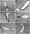
Figure 1
Figure 1
Blast-induced degenerative changes in cerebral microvessels. In panels (AD), cerebral microvessels are shown from an animal that received a single 74.5 kPa blast exposure and was sacrificed 24 h later. Panels (E,F) illustrate longitudinally cut cerebral microvessels from non-blast exposed sham controls. All microvessels in panels (AD) have lost their luminal circularity and the walls are irregular. In panel (A), a dysmorphic endothelial cell nucleus (*) is seen in the vessel lumen. In panel (D), the nucleus of a perivascular cell (arrow) with degenerative changes is indicated. Despite the destruction of the microvessels, the surrounding neuropil appears intact. Scale bar: 1 µm. Figure is reproduced from Gama Sosa et al. [34].
Figure 2
Figure 2
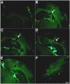
Disrupted smooth muscle layers and intraluminal astrocytic processes in blood vessels 10 months after blast exposure. Sections from rats euthanized 10 months after the last blast exposure were immunostained for GFAP (red) and α-SMA (green) with a DAPI nuclear counterstain (blue). Panels (AD) show images from the hippocampal stratum lacunosum moleculare from control (A,B) or blast-exposed (C,D) rats. Note the irregularity and vacuolation in the smooth muscle apparent with α-SMA staining in the blast-exposed animal. An arrow in (D) points to intraluminal accumulation of GFAP. Scale bar, 50 μm. Figure is reproduced from Gama Sosa et al. [35].
Figure 3
Figure 3
Reduced GFAP and fewer astroglial attachments in isolated brain vascular fractions following BINT. Panel (A) shows immunoblotting for GFAP (top panel) from five control and five blast-exposed brain-derived vascular fractions. Quantification of the blot with expression normalized to glyceraldehyde 3-phosphate dehydrogenase (GAPDH, lower panel) is shown on the right. All lanes were loaded with 10 μg of protein and contained protein from individual animals. Error bars indicate the SEM (** p < 0.01, unpaired t-test). Panel (B) shows isolated vascular fractions from control and blast-exposed brains immunostained for GFAP (red) and laminin (green) and counterstained with DAPI (blue). Arrows indicate GFAP-immunostained attachments to the isolated vasculature. Note reduced GFAP-labeled attachments in the blast-exposed vessels. All animals were euthanized 6 weeks after blast exposure. Scale bar, 25 μm. Panels (A,B) are from Gama Sosa et al. [35].
Figure 4
Figure 4
Blast-induced swelling and degeneration of astrocytic endfeet. Electron micrographs are shown from sections of the frontal motor cortex of control (A,C,F) and blast-exposed (B,D,E,G,H) rats. Animals were euthanized 6 weeks following 3 × 74.5 kPa blast exposures. Asterisks (*) indicate astrocytic endfeet, which are swollen and contain degenerating organelles in all blast-exposed microvessels. The lumens of microvessels in blast-exposed animals also appear irregular and collapsed. Perivascular astrocytes (labeled A) are visible in panels (C,E). Scale bars, 2 μm. Scale bar in panel (B) applies to panels (A,B). Scale bar in panel (C) applies to panels (CH). Scale bar in panel (D) applies only to panel (D). Figure is reproduced from Gama Sosa et al. [35].
Figure 5
Figure 5
Perivascular p-tau in astroglial processes 10 months after blast exposure. Penetrating cortical vessels from control (AC) and blast-exposed rats (DF) immunostained for AT270 (green, A,D) and GFAP (red, B,E) are shown. Asterisks in panel (A) point to the vascular lumen. The arrow in panel (D) indicates p-tau staining. Scale bar in panel (F): 20 μm. In panels (GL), a thalamic vessel is shown from a blast-exposed rat sacrificed 10 months after the last blast exposure immunostained with AT270 (green, G,J) and GFAP (red, H,K). Merged images are shown in panels (I,L). DAPI is shown in blue. Panels (J-L) show higher-power images of the vessel shown in panels (GI). Note the localization of p-tau immunostaining mostly within GFAP immunostained perivascular astroglial processes. Asterisks in panel (L) indicate endothelial cell nuclei identifiable by their elongated appearance. Scale bar in panel (L): 40 μm for panels (GI); 20 μm for panels (JL). Panels (MT) show a lack of p-tau in the vascular smooth muscle layer. Shown is a pial cortical vessel (MP) or a thalamic vessel (QT) from a blast-exposed rat sacrificed 10 months after the last blast exposure. Sections were immunostained with AT270 (green, M,Q) and α-SMA (red, N,R). DAPI is shown in blue. Merged images are shown in panels (O,S). Panels (P,T) show higher-power images of the boxed areas indicated in panels (O,S). Note the lack of co-localization of p-tau immunostaining with the α-SMA stained smooth muscle layer. Scale bar is 40 μm for (MO,QS); 10 μm for (P,T). Figures are reproduced from Dickstein et al. [33].
Figure 6
Figure 6
Altered extracellular matrix immunostaining of blast-exposed animals. Shown is collagen IV immunostaining without pepsin pretreatment of serial sections (AF) taken 1200 μm apart from a blast-exposed rat that received 3 × 74.5 kPa exposures and was sacrificed 10 months after exposure. A focal cortical lesion is indicated by arrows in panels (BD). Note the extensive lateral and rostro-caudal extent of altered collagen IV immunostaining without pepsin pretreatment. Scale bar: 750 μm. Figures are reproduced from Gama Sosa et al. [34].
Figure 7
Figure 7
Activated perivascular microglia. Shown is a luminal view of a patch of perivascular activated M1 microglia expressing MHCII. Panel (A) shows merged images; panel (B), Iba1 immunostaining (microglia, red); and panel (C), MHCII immunostaining (activated M1 microglia, green). Arrows in panel (A) show an apoptotic-activated microglial cell (green) and a degenerating perivascular microglial cell (red). Scale, 20 μm. Figure is reproduced from Gama Sosa et al. [38].
Figure 8
Figure 8
Hypothetical scenario for how early vascular injury leads to a delayed and chronic neurobehavioral phenotype. Details are discussed in the text.

References

    1. Elder G.A., Ehrlich M.E., Gandy S. Relationship of traumatic brain injury to chronic mental health problems and dementia in military veterans. Neurosci. Lett. 2019;707:134294. doi: 10.1016/j.neulet.2019.134294. - DOI - PubMed
    1. Hoge C.W., McGurk D., Thomas J.L., Cox A.L., Engel C.C., Castro C.A. Mild traumatic brain injury in U.S. Soldiers returning from Iraq. N. Engl. J. Med. 2008;358:453–463. doi: 10.1056/NEJMoa072972. - DOI - PubMed
    1. Tanielian T., Jaycox L.H. Invisible Wounds of War: Psychological and Cognitive Injuries, Their Consequences, and Services to Assist Recovery. Rand Corporation; Santa Monica, CA, USA: 2008.
    1. DePalma R.G. Combat TBI: History, Epidemiology and Injury Modes. In: Kobeissy F., editor. Brain Neurotrauma: Molecular, Neuropsychological, and Rehabilitation Aspects. CRC Press; Boca Raton, FL, USA: 2015. pp. 5–14.
    1. Elder G.A. Update on TBI and Cognitive Impairment in Military Veterans. Curr. Neurol. Neurosci. Rep. 2015;15:68. doi: 10.1007/s11910-015-0591-8. - DOI - PubMed