

Implication of lncRNA MSTRG.81401 in Hippocampal Pyroptosis Induced by P2X7 Receptor in Type 2 Diabetic Rats with Neuropathic Pain Combined with Depression
- PMID: 38256257
- PMCID: PMC10816120
- DOI: 10.3390/ijms25021186
Implication of lncRNA MSTRG.81401 in Hippocampal Pyroptosis Induced by P2X7 Receptor in Type 2 Diabetic Rats with Neuropathic Pain Combined with Depression
Abstract
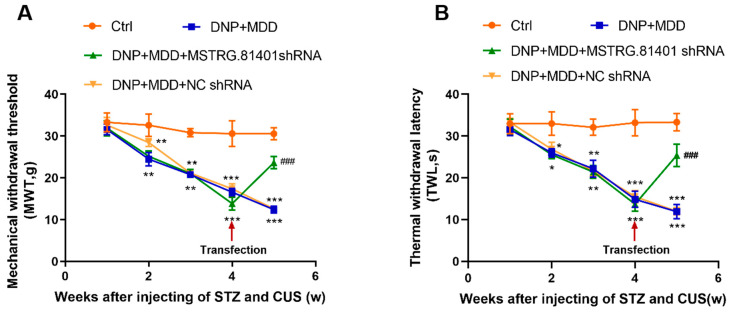
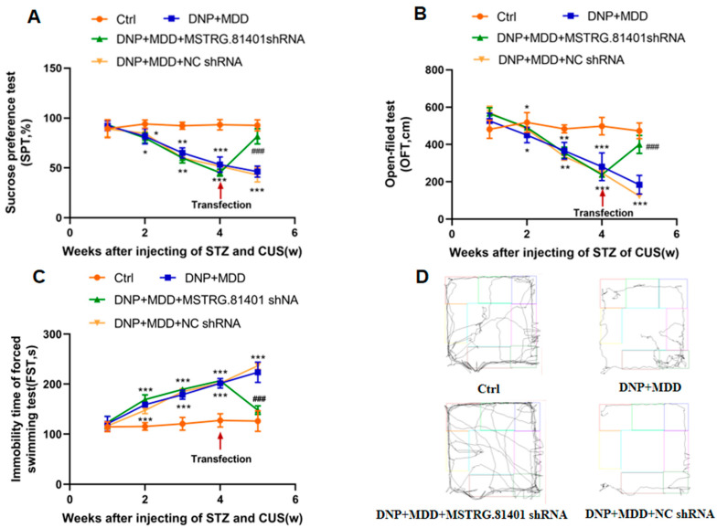
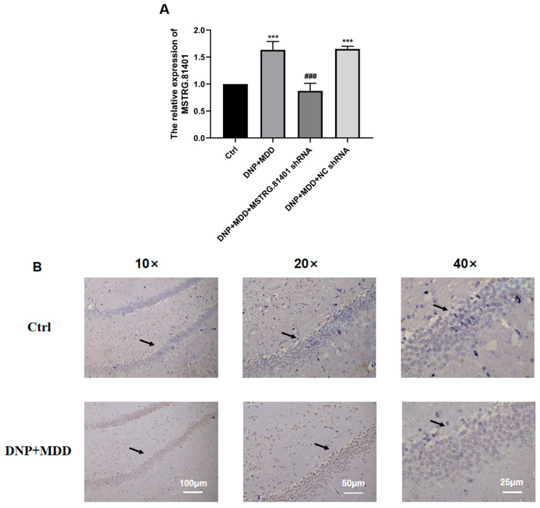
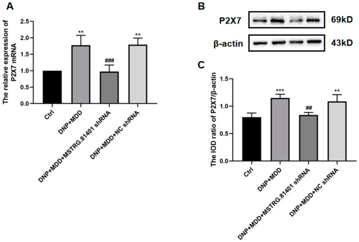
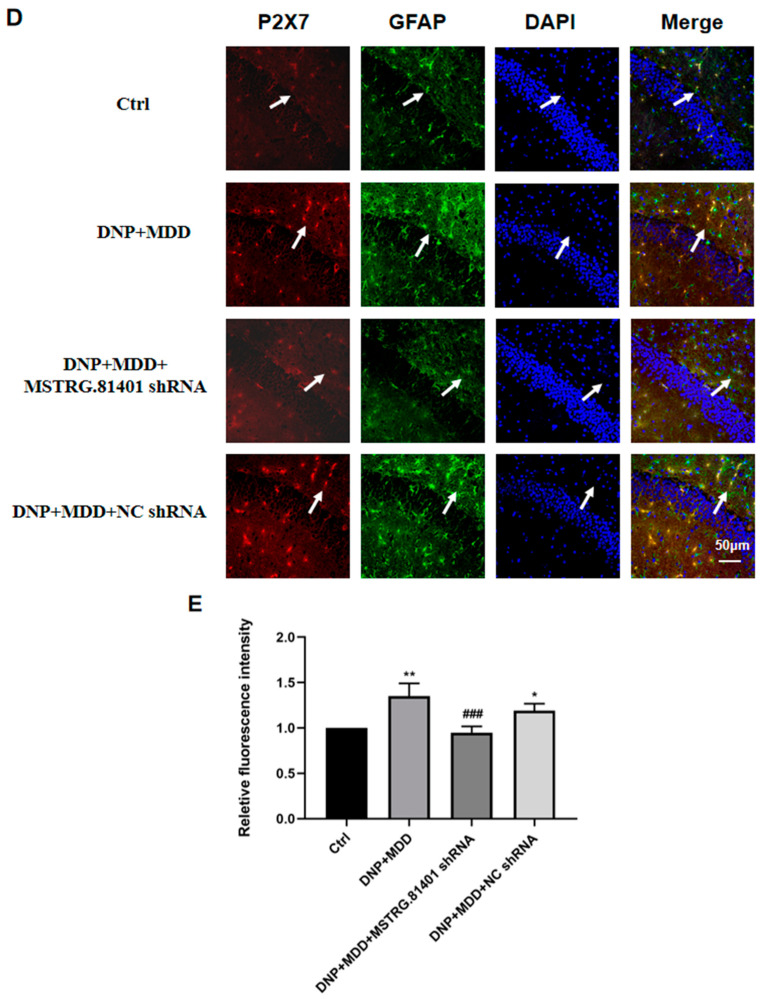
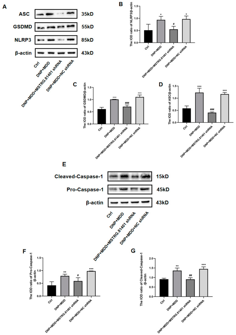
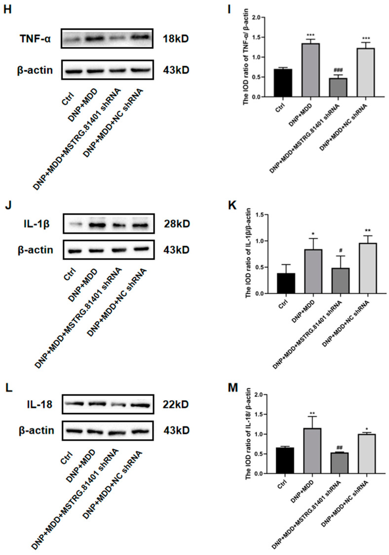
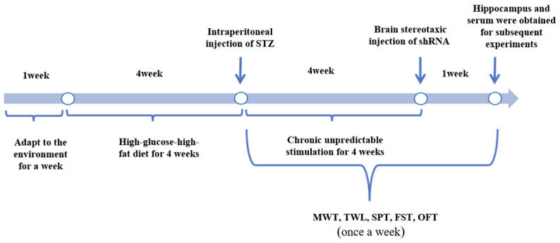
Major depressive disorder (MDD) is a common complication of diabetes and is often observed alongside diabetic neuropathic pain (DNP) as a comorbidity in diabetic patients. Long non-coding RNA (lncRNA) plays an important role in various pathophysiological processes. The P2X7 receptor is responsible for triggering inflammatory responses, such as pyroptosis, linked to pain and depression. The aim of this study was to investigate the effect of lncRNA MSTRG.81401 on hippocampal pyroptosis induced by the P2X7 receptor in diabetic rats with DNP combined with MDD (DNP + MDD). Our results showed that the expression of lncRNA MSTRG.81401 was significantly elevated in the hippocampus of DNP + MDD rats compared with the control group. Following the administration of shRNA targeting lncRNA MSTRG.81401, a notable elevation in mechanical and thermal pain thresholds was observed in rats with comorbid DNP and MDD. Additionally, significant improvements in depression-like behaviors were evident in the open-field test (OFT), sucrose preference test (SPT), and forced swim test (FST). In the DNP + MDD rats, elevated levels in hippocampal P2X7 receptor mRNA and protein were observed, along with increased co-expression of P2X7 and the astrocytic marker glial fibrillary acidic protein (GFAP). Meanwhile, in DNP + MDD rats, the heightened mRNA expression of NOD-like receptor protein 3 (NLRP3), apoptosis-associated speck-like protein (ASC), pyroptosis-related protein Gasdermin D (GSDMD), caspase-1, IL-1β, IL-18, and TNF-α was detected, in addition to increased serum levels of IL-1β, IL-18 and TNF-α. After shRNA treatment with lncRNA MSTRG.81401, the above abnormal changes in indicators for pyroptosis and inflammation were improved. Therefore, our study demonstrates that shRNA of lncRNA MSTRG.81401 can alleviate the pain and depression-like behaviors in diabetic rats associated with the comorbidity of DNP and MDD by inhibiting the hippocampal P2X7 receptor-mediated pyroptosis pathway and pro-inflammatory responses. This suggests that the P2X7R/NLRP3/caspase-1 implicated pyroptosis and inflammatory scenario may serve as a potential target for the management of comorbid DNP and MDD in diabetes.
Keywords: P2X7 receptor; diabetic neuropathologic pain; hippocampus; lncRNA; major depressive disorder; pyroptosis.
Conflict of interest statement
The authors declare that they have no financial or other conflicts of interest in association with this work.
Figures
Similar articles
-
Long non-coding RNA MSTRG.81401 short hairpin RNA relieves diabetic neuropathic pain and behaviors of depression by inhibiting P2X4 receptor expression in type 2 diabetic rats.Purinergic Signal. 2023 Mar;19(1):123-133. doi: 10.1007/s11302-021-09828-0. Epub 2022 Jan 13. Purinergic Signal. 2023. PMID: 35022948 Free PMC article.
-
LncRNA NONRATT021972 siRNA regulates neuropathic pain behaviors in type 2 diabetic rats through the P2X7 receptor in dorsal root ganglia.Mol Brain. 2016 Apr 23;9:44. doi: 10.1186/s13041-016-0226-2. Mol Brain. 2016. PMID: 27107575 Free PMC article.
-
Effects of palmatine on rats with comorbidity of diabetic neuropathic pain and depression.Brain Res Bull. 2018 May;139:56-66. doi: 10.1016/j.brainresbull.2018.02.005. Epub 2018 Feb 7. Brain Res Bull. 2018. PMID: 29427595
-
Genetic and Epigenetic Regulation of the Innate Immune Response to Gout.Immunol Invest. 2023 Apr;52(3):364-397. doi: 10.1080/08820139.2023.2168554. Epub 2023 Feb 6. Immunol Invest. 2023. PMID: 36745138 Review.
-
P2X7 Receptor as a Potential Target for Major Depressive Disorder.Curr Drug Targets. 2021;22(10):1108-1120. doi: 10.2174/1389450122666210120141908. Curr Drug Targets. 2021. PMID: 33494675 Review.
Cited by
-
Pyroptosis in health and disease: mechanisms, regulation and clinical perspective.Signal Transduct Target Ther. 2024 Sep 20;9(1):245. doi: 10.1038/s41392-024-01958-2. Signal Transduct Target Ther. 2024. PMID: 39300122 Free PMC article. Review.
-
Different Types of Cell Death in Diabetic Neuropathy: A Focus on Mechanisms and Therapeutic Strategies.Int J Mol Sci. 2024 Jul 25;25(15):8126. doi: 10.3390/ijms25158126. Int J Mol Sci. 2024. PMID: 39125694 Free PMC article. Review.
-
mtDNA-cGAS-STING axis-dependent NLRP3 inflammasome activation contributes to postoperative cognitive dysfunction induced by sevoflurane in mice.Int J Biol Sci. 2024 Mar 3;20(5):1927-1946. doi: 10.7150/ijbs.91543. eCollection 2024. Int J Biol Sci. 2024. PMID: 38481801 Free PMC article.
-
The Association and Prognostic Implications of Long Non-Coding RNAs in Major Psychiatric Disorders, Alzheimer's Diseases and Parkinson's Diseases: A Systematic Review.Int J Mol Sci. 2024 Oct 12;25(20):10995. doi: 10.3390/ijms252010995. Int J Mol Sci. 2024. PMID: 39456775 Free PMC article.
-
Comprehension of gut microbiota and microRNAs may contribute to the development of innovative treatment tactics against metabolic disorders and psychiatric disorders.Int J Physiol Pathophysiol Pharmacol. 2024 Dec 25;16(6):111-125. doi: 10.62347/WAZH2090. eCollection 2024. Int J Physiol Pathophysiol Pharmacol. 2024. PMID: 39850247 Free PMC article. Review.
References
MeSH terms
Substances
Grants and funding
LinkOut - more resources
Full Text Sources
Medical
Miscellaneous
