. 2024 Jan 10;60(1):126.
doi: 10.3390/medicina60010126.

Redefining the Axillary Aesthetic: Surgical Management of Axillary Tissue Hypertrophy

Affiliations

Redefining the Axillary Aesthetic: Surgical Management of Axillary Tissue Hypertrophy

Neil Tanna et al. Medicina (Kaunas). .

Abstract

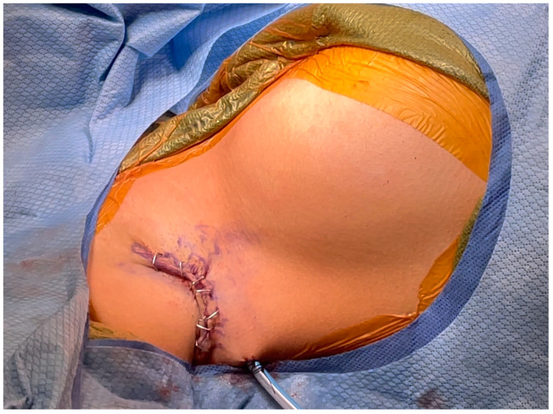
Background and Objectives: Axillary tissue hypertrophy consists of ectopic breast tissue and occurs in up to six percent of women. Women complain of pain, interference with activity, and dissatisfaction with appearance. While it is recommended that accessory breast tissue be removed via surgical excision, there is lack of consensus on the best technique for the surgical management of axillary tissue hypertrophy. In this study, the senior authors (BC and NT) review outcomes and complications as they pertain to the surgical treatment of axillary tissue hypertrophy and axillary contouring. Materials and Methods: A retrospective review of all patients (n = 35), from two separate institutions, who presented with axillary tissue hypertrophy between December 2019 and August 2021 was conducted. All patients underwent a technique that included direct crescentic dermato-lipectomy and glandular excision with axillary crease obliteration. Tissue was sent for histological analysis after removal. During a six-month follow-up period, all patient outcomes were recorded. Results: The authors treated 35 women with axillary tissue hypertrophy. All patients complained of aesthetic deformity with significant discomfort leading to the desire for surgery. Histologically, all specimens contained benign breast and adipose tissue. Hypertrophic scarring, seroma, and axillary cording were noted complications. Conclusions: Detailed is the surgical management and optimal technique that can be used to treat both adipose and fibroglandular axillary tissue hypertrophy while simultaneously providing a favorable axillary aesthetic.

Keywords: aesthetic surgery; axillary tissue; breast surgery.


Conflict of interest statement

The authors declare no conflicts of interest.

Figures

Figure 1
Figure 1
Axillary tissue hypertrophy. Axillary ectopic breast tissue consists of ectopic mammary tissue that develops outside the normal breast region. Most commonly it is found in the axilla but can occur anywhere along the embryonic mammary streak.
Figure 2
Figure 2
Preoperative demarcation. The patient is marked with the arm in 45 degrees abduction. Within the axillary fossa, a crescentic area of excision is marked, including the overlying skin and the underlying adipose and glandular tissue (solid line). The area of dissection that will be beveled outwards is also denoted (cloud line). Any axillary folds or creases, if present, are also demarcated.
Figure 3
Figure 3
Axillary tissue incision. The skin is incised along the marked crescentric area within the axillary fossa. The posterior incision dissection is performed perpendicular, straight down, through the subcutaneous fat.
Figure 4
Figure 4
Tissue beveling and axillary crease obliteration. After the skin is incised, the dissection is beveled outward anteriorly. This creates a subcutaneous flap anteriorly. At all locations, the dissection is performed down to, but not violating, the clavipectoral fascia. Anteriorly, any axillary creases or folds are obliterated by the scoring of the subcutaneous tissue.
Figure 5
Figure 5
Axillary tissue excision. The excised axillary tissue and resulting defect are shown. Removing the area of excess tissue results in dead space that may be prone to serous fluid accumulation. To prevent this, a 15-French Blake drain is placed posteriorly through a separate stab incision.
Figure 6
Figure 6
Meticulous skin alignment for closure. The skin edges are meticulously aligned at the anterior and posterior edges with redundancy kept in the middle. The incisions are then closed in two layers.
Figure 7
Figure 7
Postoperative result. Careful demarcation and incision ensure that the resulting scar is within the axillary fossa. Therefore, postoperatively, the scar will not be visible with the patient supine or arm adducted.
Figure 8
Figure 8
Before and after. Preoperative and postoperative comparison of a patient with axillary tissue hypertrophy.

References

    1. Schoenwolf G.C., Bleyl S.B., Brauer P.R., Francis-West P.H. Development of the skin and its derivatives. In: Larsen W.J., editor. Human Embryology. 4th ed. Churchill Livingstone; Philadelphia, PA, USA: 2009. pp. 193–216.
    1. Hwang S.B., Choi B.S., Byun G.Y., Koo B.H., Lee S.R. Accessory Axillary Breast Excision with Liposuction Using Minimal Incision: A Preliminary Report. Aesthetic Plast. Surg. 2016;41:10–18. doi: 10.1007/s00266-016-0729-3. - DOI - PubMed
    1. Greer K.E. Accessory axillary breast tissue. Arch. Dermatol. 1974;109:88–89. doi: 10.1001/archderm.1974.01630010064017. - DOI - PubMed
    1. Youn H.J., Jung S.H. Accessory Breast Carcinoma. Breast Care. 2009;4:104–106. doi: 10.1159/000210638. - DOI - PMC - PubMed
    1. Sahu S.K., Husain M., Sachan P.K. Bilateral accessory breast. Internet J. Surg. 2008