

Thyroid Imaging Reporting and Data Systems: Applicability of the "Taller than Wide" Criterium in Primary/Secondary Care Units and the Role of Thyroid Scintigraphy
- PMID: 38256648
- PMCID: PMC10816136
- DOI: 10.3390/jcm13020514
Thyroid Imaging Reporting and Data Systems: Applicability of the "Taller than Wide" Criterium in Primary/Secondary Care Units and the Role of Thyroid Scintigraphy
Abstract
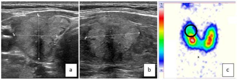
Background: To examine the applicability of the "taller than wide" (ttw) criterium for risk assessment of thyroid nodules (TNs) in primary/secondary care units and the role of thyroid scintigraphy therein.
Methods: German bicenter study performed in a setting of primary/secondary care. Patient recruitment and analysis in center A was conducted in a prospective manner. In center B, patient data were retrieved from a database that was originally generated by prospective data collection. TNs were assessed by ultrasound and thyroid scans, mostly fine needle biopsy and occasionally surgery and others. In center A, only patients who presented for the first time were included. The inclusion criterion was any TN ≥ 10 mm that had at least the following two sonographic risk features: solidity and a ttw shape. In center B, consecutive patients who had at least ttw and hypofunctioning nodules ≥ 10 mm were retrieved from the above-mentioned database. The risk of malignancy was determined according to a mixed reference standard and compared with literature data.
Results: In center A, 223 patients with 259 TNs were included into the study. For further analysis, 200 nodules with a reference standard were available. The overall malignancy rate was 2.5% (upper limit of the 95% CI: 5.1%). After the exclusion of scintigraphically hyperfunctioning nodules, the malignancy rate increased slightly to 2.8% (upper limit of the 95% CI: 5.7%). Malignant nodules exhibited sonographic risk features additional to solidity and ttw shape more often than benign ones. In addition to the exclusion of hyperfunctioning nodules, when considering only nodules without additional US risk features, i.e., exclusively solid and ttw-nodules, the malignancy rate decreased to 0.9% (upper limit 95% CI: 3.7%). In center B, from 58 patients, 58 ttw and hypofunctioning TNs on thyroid scans with a reference standard were available. Malignant nodules from center B were always solid and hypoechoic. The overall malignancy rate of hypofunctioning and ttw nodules was 21%, with the lower limit of the 95% CI (one-sided) being 12%.
Conclusions: In primary/secondary care units, the lowest TIRADS categories for indicating FNB, e.g., applying one out of five sonographic risk features, may not be appropriate owing to the much lower a priori malignancy risk in TNs compared to tertiary/quaternary care units. Even the combination of two sonographic risk features, "solidity" and "ttw", may only be appropriate in a limited fashion. In contrast, the preselection of TNs according to hypofunctioning findings on thyroid scans clearly warranted FNB, even when applying only one sonographic risk criterion ("ttw"). For this reason, thyroid scans in TNs may not only be indicated to rule out hyperfunctioning nodules from FNB but also to rule in hypofunctioning ones.
Keywords: Thyroid Imaging Reporting and Data Systems; fine needle biopsy; risk of malignancy; taller than wide; thyroid nodule; thyroid scintigraphy.
Conflict of interest statement
The authors declare no conflict of interest.
Figures

Similar articles
-
Introducing a Pole Concept for Nodule Growth in the Thyroid Gland: Taller-than-Wide Shape, Frequency, Location and Risk of Malignancy of Thyroid Nodules in an Area with Iodine Deficiency.J Clin Med. 2022 May 1;11(9):2549. doi: 10.3390/jcm11092549. J Clin Med. 2022. PMID: 35566675 Free PMC article.
-
Distribution of Functional Status of Thyroid Nodules and Malignancy Rates of Hyperfunctioning and Hypofunctioning Thyroid Nodules in Germany.Nuklearmedizin. 2022 Oct;61(5):376-384. doi: 10.1055/a-1856-4052. Epub 2022 Aug 2. Nuklearmedizin. 2022. PMID: 35917825 English.
-
Which ultrasound image plane is appropriate for evaluating the taller-than-wide sign in the risk stratification of thyroid nodules?Eur Radiol. 2021 Oct;31(10):7605-7613. doi: 10.1007/s00330-021-07936-4. Epub 2021 Apr 14. Eur Radiol. 2021. PMID: 33855586
-
Correlation between ultrasonographic and cytologic features of thyroid nodules: a single-center cross-sectional study.J Med Life. 2024 Jun;17(6):593-600. doi: 10.25122/jml-2024-0038. J Med Life. 2024. PMID: 39296443 Free PMC article.
-
Diagnostic performance of adult-based ATA and ACR-TIRADS ultrasound risk stratification systems in pediatric thyroid nodules: a systematic review and meta-analysis.Eur Radiol. 2021 Oct;31(10):7450-7463. doi: 10.1007/s00330-021-07908-8. Epub 2021 Apr 17. Eur Radiol. 2021. PMID: 33864505
References
-
- Kwak J.Y., Han K.H., Yoon J.H., Moon H.J., Son E.J., Park S.H., Jung H.K., Choi J.S., Kim B.M., Kim E.K. Thyroid imaging reporting and data system for US features of nodules: A step in establishing better stratification of cancer risk. Radiology. 2011;260:892–899. doi: 10.1148/radiol.11110206. - DOI - PubMed
-
- Shin J.H., Baek J.H., Chung J., Ha E.J., Kim J.-H., Lee Y.H., Lim H.K., Moon W.-J., Na D.G., Park J.S., et al. Ultrasonography Diagnosis and Imaging-Based Management of Thyroid Nodules: Revised Korean Society of Thyroid Radiology Consensus Statement and Recommendations. Korean J. Radiol. 2016;17:370–395. doi: 10.3348/kjr.2016.17.3.370. - DOI - PMC - PubMed
-
- Tessler F.N., Middleton W.D., Grant E.G., Hoang J.K., Berland L.L., Teefey S.A., Cronan J.J., Beland M.D., Desser T.S., Frates M.C., et al. ACR Thyroid Imaging, Reporting and Data System (TI-RADS): White Paper of the ACR TI-RADS Committee. J. Am. Coll. Radiol. 2017;14:587–595. doi: 10.1016/j.jacr.2017.01.046. - DOI - PubMed
LinkOut - more resources
Full Text Sources
