

Magnesium Hydroxide as a Versatile Nanofiller for 3D-Printed PLA Bone Scaffolds
- PMID: 38256997
- PMCID: PMC10820754
- DOI: 10.3390/polym16020198
Magnesium Hydroxide as a Versatile Nanofiller for 3D-Printed PLA Bone Scaffolds
Abstract
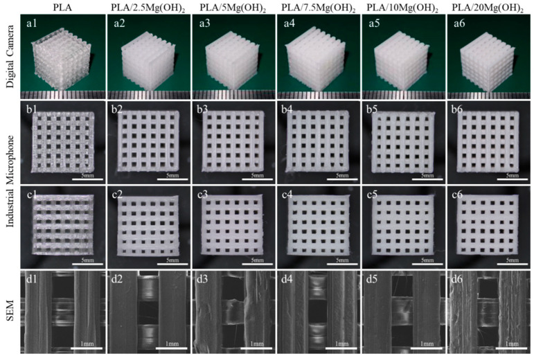
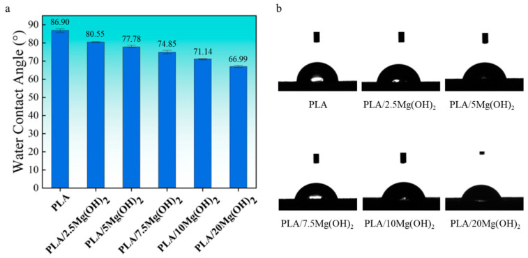
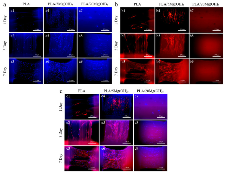
Polylactic acid (PLA) has attracted much attention in bone tissue engineering due to its good biocompatibility and processability, but it still faces problems such as a slow degradation rate, acidic degradation product, weak biomineralization ability, and poor cell response, which limits its wider application in developing bone scaffolds. In this study, Mg(OH)2 nanoparticles were employed as a versatile nanofiller for developing PLA/Mg(OH)2 composite bone scaffolds using fused deposition modeling (FDM) 3D printing technology, and its mechanical, degradation, and biological properties were evaluated. The mechanical tests revealed that a 5 wt% addition of Mg(OH)2 improved the tensile and compressive strengths of the PLA scaffold by 20.50% and 63.97%, respectively. The soaking experiment in phosphate buffered solution (PBS) revealed that the alkaline degradation products of Mg(OH)2 neutralized the acidic degradation products of PLA, thus accelerating the degradation of PLA. The weight loss rate of the PLA/20Mg(OH)2 scaffold (15.40%) was significantly higher than that of PLA (0.15%) on day 28. Meanwhile, the composite scaffolds showed long-term Mg2+ release for more than 28 days. The simulated body fluid (SBF) immersion experiment indicated that Mg(OH)2 promoted the deposition of apatite and improved the biomineralization of PLA scaffolds. The cell culture of bone marrow mesenchymal stem cells (BMSCs) indicated that adding 5 wt% Mg(OH)2 effectively improved cell responses, including adhesion, proliferation, and osteogenic differentiation, due to the release of Mg2+. This study suggests that Mg(OH)2 can simultaneously address various issues related to polymer scaffolds, including degradation, mechanical properties, and cell interaction, having promising applications in tissue engineering.
Keywords: biological properties; bone scaffold; degradation properties; fused deposition modeling (FDM); magnesium hydroxide (Mg(OH)2); mechanical properties; polylactic acid (PLA).
Conflict of interest statement
The authors declare no conflicts of interest.
Figures


Similar articles
-
A natural biomineral for enhancing the biomineralization and cell response of 3D printed polylactic acid bone scaffolds.Int J Biol Macromol. 2023 Jul 1;242(Pt 1):124728. doi: 10.1016/j.ijbiomac.2023.124728. Epub 2023 May 5. Int J Biol Macromol. 2023. PMID: 37150372
-
Inorganic whiskers containing alkaline and bioactive ions enhance the comprehensive properties of 3D-printed biopolymer bone scaffold.Biomed Mater. 2025 Aug 12;20(5). doi: 10.1088/1748-605X/adf619. Biomed Mater. 2025. PMID: 40738142
-
3D printed TPMS structural PLA/GO scaffold: Process parameter optimization, porous structure, mechanical and biological properties.J Mech Behav Biomed Mater. 2023 Jun;142:105848. doi: 10.1016/j.jmbbm.2023.105848. Epub 2023 Apr 18. J Mech Behav Biomed Mater. 2023. PMID: 37099921
-
Fused Deposition Modeling Printed PLA/Nano β-TCP Composite Bone Tissue Engineering Scaffolds for Promoting Osteogenic Induction Function.Int J Nanomedicine. 2023 Oct 17;18:5815-5830. doi: 10.2147/IJN.S416098. eCollection 2023. Int J Nanomedicine. 2023. PMID: 37869064 Free PMC article.
-
On the Fused Deposition Modelling of Personalised Bio-Scaffolds: Materials, Design, and Manufacturing Aspects.Bioengineering (Basel). 2024 Jul 31;11(8):769. doi: 10.3390/bioengineering11080769. Bioengineering (Basel). 2024. PMID: 39199727 Free PMC article. Review.
Cited by
-
Effect of Annealing on the Mechanical Properties of Composites of PLA Mixed with Mg and with HA.Polymers (Basel). 2025 Apr 28;17(9):1207. doi: 10.3390/polym17091207. Polymers (Basel). 2025. PMID: 40362991 Free PMC article.
-
Improvement in Crystallization, Thermal, and Mechanical Properties of Flexible Poly(L-lactide)-b-poly(ethylene glycol)-b-poly(L-lactide) Bioplastic with Zinc Phenylphosphate.Polymers (Basel). 2024 Apr 3;16(7):975. doi: 10.3390/polym16070975. Polymers (Basel). 2024. PMID: 38611233 Free PMC article.
-
Three-Dimensional-Printed Biomimetic Scaffolds for Investigating Osteoblast-Like Cell Interactions in Simulated Microgravity: An In Vitro Platform for Bone Tissue Engineering Research.J Funct Biomater. 2025 Jul 24;16(8):271. doi: 10.3390/jfb16080271. J Funct Biomater. 2025. PMID: 40863291 Free PMC article.
-
Development and Application of Polymer Scaffolds.Polymers (Basel). 2025 Aug 21;17(16):2260. doi: 10.3390/polym17162260. Polymers (Basel). 2025. PMID: 40871206 Free PMC article.
-
Advances in biomaterials for osteonecrosis treatment.Front Pharmacol. 2025 May 21;16:1559810. doi: 10.3389/fphar.2025.1559810. eCollection 2025. Front Pharmacol. 2025. PMID: 40469973 Free PMC article. Review.
References
-
- Collins M.N., Ren G., Young K., Pina S., Reis R.L., Oliveira J.M. Scaffold Fabrication Technologies and Structure/Function Properties in Bone Tissue Engineering. Adv. Funct. Mater. 2021;31:2010609. doi: 10.1002/adfm.202010609. - DOI
-
- Yang J., Wang H., Huang W., Peng K., Shi R., Tian W., Lin L., Yuan J., Yao W., Ma X., et al. A Natural Polymer-Based Hydrogel with Shape Controllability and High Toughness and Its Application to Efficient Osteochondral Regeneration. Mater. Horiz. 2023;10:3797–3806. doi: 10.1039/D3MH00544E. - DOI - PubMed
-
- Krobot S., Melcova V., Mencik P., Kontarova S., Rampichova M., Hedvicakova V., Mojzisova E., Baco A., Prikryl R. Poly(3-Hydroxybutyrate) (PHB) and Polycaprolactone (PCL) Based Blends for Tissue Engineering and Bone Medical Applications Processed by FDM 3D Printing. Polymers. 2023;15:2404. doi: 10.3390/polym15102404. - DOI - PMC - PubMed
Grants and funding
LinkOut - more resources
Full Text Sources
