Polycaprolactone Nanofibers Functionalized by Fibronectin/Gentamicin and Implanted Silver for Enhanced Antibacterial Properties, Cell Adhesion, and Proliferation
- PMID: 38257060
- PMCID: PMC10819432
- DOI: 10.3390/polym16020261
Polycaprolactone Nanofibers Functionalized by Fibronectin/Gentamicin and Implanted Silver for Enhanced Antibacterial Properties, Cell Adhesion, and Proliferation
Abstract
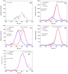
Novel nanomaterials used for wound healing should have many beneficial properties, including high biological and antibacterial activity. Immobilization of proteins can stimulate cell migration and viability, and implanted Ag ions provide an antimicrobial effect. However, the ion implantation method, often used to introduce a bactericidal element into the surface, can lead to the degradation of vital proteins. To analyze the surface structure of nanofibers coated with a layer of plasma COOH polymer, fibronectin/gentamicin, and implanted with Ag ions, a new X-ray photoelectron spectroscopy (XPS) fitting method is used for the first time, allowing for a quantitative assessment of surface biomolecules. The results demonstrated noticeable changes in the composition of fibronectin- and gentamicin-modified nanofibers upon the introduction of Ag ions. Approximately 60% of the surface chemistry has changed, mainly due to an increase in hydrocarbon content and the introduction of up to 0.3 at.% Ag. Despite the significant degradation of fibronectin molecules, the biological activity of Ag-implanted nanofibers remained high, which is explained by the positive effect of Ag ions inducing the generation of reactive oxygen species. The PCL nanofibers with immobilized gentamicin and implanted silver ions exhibited very significant antipathogen activity to a wide range of Gram-positive and Gram-negative strains. Thus, the results of this work not only make a significant contribution to the development of new hybrid fiber materials for wound dressings but also demonstrate the capabilities of a new XPS fitting methodology for quantitative analysis of surface-related proteins and antibiotics.
Keywords: Immobilization; PCL nanofibers; XPS; fibronectin; plasma; silver; surface functionalization.
Conflict of interest statement
The authors declare no conflicts of interest.
Figures


Similar articles
-
Plasma-Coated Polycaprolactone Nanofibers with Covalently Bonded Platelet-Rich Plasma Enhance Adhesion and Growth of Human Fibroblasts.Nanomaterials (Basel). 2019 Apr 19;9(4):637. doi: 10.3390/nano9040637. Nanomaterials (Basel). 2019. PMID: 31010178 Free PMC article.
-
Self-Sanitizing Polycaprolactone Electrospun Nanofiber Membrane with Ag Nanoparticles.J Funct Biomater. 2023 Jun 25;14(7):336. doi: 10.3390/jfb14070336. J Funct Biomater. 2023. PMID: 37504830 Free PMC article.
-
Immobilization of Platelet-Rich Plasma onto COOH Plasma-Coated PCL Nanofibers Boost Viability and Proliferation of Human Mesenchymal Stem Cells.Polymers (Basel). 2017 Dec 20;9(12):736. doi: 10.3390/polym9120736. Polymers (Basel). 2017. PMID: 30966035 Free PMC article.
-
Polycaprolactone based electrospun matrices loaded with Ag/hydroxyapatite as wound dressings: Morphology, cell adhesion, and antibacterial activity.Int J Pharm. 2021 Jan 25;593:120143. doi: 10.1016/j.ijpharm.2020.120143. Epub 2020 Dec 3. Int J Pharm. 2021. PMID: 33279712
-
In vitro antimicrobial and anticancer properties of TiO2 blow-spun nanofibers containing silver nanoparticles.Mater Sci Eng C Mater Biol Appl. 2019 Nov;104:109876. doi: 10.1016/j.msec.2019.109876. Epub 2019 Jun 8. Mater Sci Eng C Mater Biol Appl. 2019. PMID: 31500007
Cited by
-
Hydrogel Containing Propolis: Physical Characterization and Evaluation of Biological Activities for Potential Use in the Treatment of Skin Lesions.Pharmaceuticals (Basel). 2024 Oct 20;17(10):1400. doi: 10.3390/ph17101400. Pharmaceuticals (Basel). 2024. PMID: 39459039 Free PMC article.
References
-
- Karuppuswamy P., Venugopal J.R., Navaneethan B., Laiva A.L., Sridhar S., Ramakrishna S. Functionalized hybrid nanofibers to mimic native ECM for tissue engineering applications. Appl. Surf. Sci. 2014;322:162–168. doi: 10.1016/j.apsusc.2014.10.074. - DOI
-
- Prosecká E., Rampichová M., Litvinec A., Tonar Z., Králíčková M., Vojtová L., Kochová P., Plencner M., Buzgo M., Míčková A., et al. Collagen/hydroxyapatite scaffold enriched with polycaprolactone nanofibers, thrombocyte-rich solution and mesenchymal stem cells promotes regeneration in large bone defect in vivo. J. Biomed. Mater. Res. Part A. 2015;103:671–682. doi: 10.1002/jbm.a.35216. - DOI - PubMed
Grants and funding
LinkOut - more resources
Full Text Sources

