. 2024 Jan 6;29(2):298.
doi: 10.3390/molecules29020298.

Chemical Constituents of Halophyte Suaeda glauca and Their Therapeutic Potential for Hair Loss

Affiliations

Chemical Constituents of Halophyte Suaeda glauca and Their Therapeutic Potential for Hair Loss

Yun-Na Kim et al. Molecules. .

Abstract

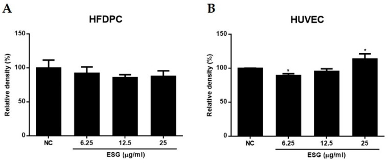
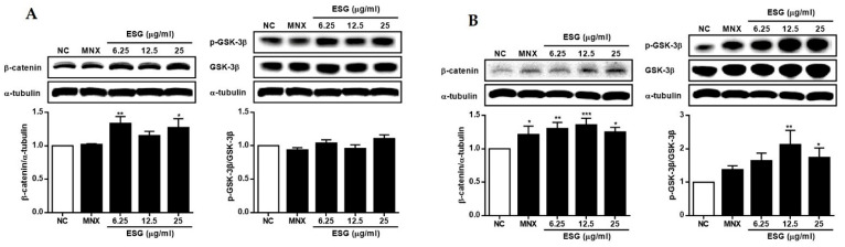
Suaeda glauca, a halophyte in the Amaranthaceae family, exhibits remarkable resilience to high salt and alkali stresses despite the absence of salt glands or vesicles in its leaves. While there is growing pharmacological interest in S. glauca, research on its secondary metabolites remains limited. In this study, chemical constituents of the aerial parts of S. glauca were identified using 1D- and 2D-NMR experiments, and its biological activity concerning hair loss was newly reported. Eight compounds, including alkaloids (1~3), flavonoids (4~6), and phenolics (7 and 8), were isolated. The compounds, except the flavonoids, were isolated for the first time from S. glauca. In the HPLC chromatogram, quercetin-3-O-β-d-glucoside, kaempferol-3-O-β-d-glucoside, and kaempferol were identified as major constituents in the extract of S. glauca. Additionally, the therapeutic potential of the extract of S. glauca and the isolated compounds 1~8 on the expressions of VEGF and IGF-1, as well as the regulation of Wnt/β-catenin signaling, were evaluated in human follicle dermal papilla cells (HFDPCs) and human umbilical vein endothelial cells (HUVECs). Among the eight compounds, compound 4 was the most potent in terms of increasing the expression of VEGF and IGF-1 and the regulation of Wnt/β-catenin. These findings suggest that S. glauca extract and its compounds are potential new candidates for preventing or treating hair loss.

Keywords: Suaeda glauca; alopecia; compounds; dermal papilla cells; hair loss; halophyte.


Conflict of interest statement

The authors declare no conflicts of interest.

Figures

Figure 1
Figure 1
Chemical structures of 1~8 isolated from the aerial parts of S. glauca.
Figure 2
Figure 2
HPLC chromatograms of the extract of S. glauca (ESG) (A) and mixture of standard compounds: kaempferol (4), kaempferol-3-O-β-d-glucoside (5), quercetin-3-O-β-d-glucoside (6) (B), and UV spectra of each standard (C).
Figure 3
Figure 3
The effects of the extract of S. glauca (ESG) on cell viability of human follicle dermal papilla cells (HFDPCs) (A) and human umbilical vein endothelial cells (HUVECs) (B). Cells were treated with ESG (6.25, 12.5, and 25 μg/mL) for 24 h, and cell viability was determined by MTT assay. Results are presented as the mean ± S.D. of triplicate experiments; * p < 0.05 compared to non-treated control (NC).
Figure 4
Figure 4
The effects of the extract of S. glauca (ESG) on the expressions of VEGF and IGF-1 in human follicle dermal papilla cells (HFDPCs) (A) and in human umbilical vein endothelial cells (HUVECs) (B). Cells were treated with ESG (6.25, 12.5 and 25 μg/mL) or MNX (10 μM, a positive control) for 24 h. The expression levels of each protein in cells were analyzed by Western blot. Results are presented as the mean ± S.D. of triplicate experiments; * p < 0.05, ** p < 0.01, *** p < 0.001 compared to NC. VEGF: vascular endothelial growth factor, IGF-1: insulin-like growth factor, MNX: minoxidil, NC: non-treated control.
Figure 5
Figure 5
The effects of the extract of S. glauca (ESG) on the expressions of β-catenin and the phosphorylation of GSK-3β proteins in human follicle dermal papilla cells (HFDPCs) (A) and in human umbilical vein endothelial cells (HUVECs) (B). Cells were treated with ESG (6.25, 12.5, and 25 μg/mL) or MNX (10 μM, a positive control) for 24 h. The expression levels of each protein in cells were analyzed by western blot. Results are presented as the mean ± S.D. of triplicate experiments; * p < 0.05, ** p < 0.01, *** p < 0.001 compared to NC. GSK-3β: glycogen synthase kinase-3 beta, MNX: minoxidil, NC: non-treated control.
Figure 6
Figure 6
The toxicities of 1~8 isolated from S. glauca against human follicle dermal papilla cells (HFDPCs) (A) and human umbilical vein endothelial cells (HUVECs) (B). Cells were treated with each compound (0.1, 1.0, 5.0, and 10.0 μM) for 24 h, and cell viability was determined by MTT assay. Results are presented as the mean ± S.D. of triplicate experiments.
Figure 7
Figure 7
The effects of compounds 18, isolated from S. glauca, on the expressions of VEGF (A), IGF-1 (B), β-catenin (C), and the phosphorylation of GSK-3β (D) proteins in human follicle dermal papilla cells (HFDPCs). Cells were treated with compounds (10 μM) or MNX (10 μM, a positive control) for 24 h. The expression levels of each protein in cells were analyzed by Western blot. Results are presented as the mean ± S.D. of triplicate experiments; ** p < 0.01 and *** p < 0.001 compared to NC. VEGF: vascular endothelial growth factor, IGF-1: insulin-like growth factor, GSK-3β: glycogen synthase kinase-3 beta, MNX: minoxidil, NC: non-treated control.
Figure 8
Figure 8
The effects of compounds 18 isolated from S.glauca on the expressions of IGF-1 (A), β-catenin (B) and the phosphorylation of GSK-3β (C) proteins in human umbilical vein endothelial cells (HUVECs). Cells were treated with compounds (10 μM) or MNX (10 μM, a positive control) for 24 h. The expression levels of each protein in cells were analyzed by Western blot. Results are presented as the mean ± S.D. of triplicate experiments; * p < 0.05, ** p < 0.01, *** p < 0.001 compared to NC. IGF-1: insulin-like growth factor, GSK-3β: glycogen synthase kinase-3 beta, MNX: minoxidil, NC: non-treated control.

References

    1. An R.B., Sohn D.H., Jeong G.S., Kim Y.C. In vitro hepatoprotective compounds from Suaeda glauca. Arch. Pharm. Res. 2008;31:594–597. doi: 10.1007/s12272-001-1198-1. - DOI - PubMed
    1. Sun H.X., Zhong R.Z., Liu H.W., Wang M.L., Sun J.Y., Zhou D.W. Meat quality, fatty acid composition of tissue and gastrointestinal content, and antioxidant status of lamb fed seed of a halophyte (Suaeda glauca) Meat Sci. 2015;100:10–16. doi: 10.1016/j.meatsci.2014.09.005. - DOI - PubMed
    1. Yang C., Shi D., Wang D. Comparative effects of salt and alkali stresses on growth, osmotic adjustment and ionic balance of an alkali-resistant halophyte Suaeda glauca (Bge.) Plant Growth Reg. 2008;56:179–190. doi: 10.1007/s10725-008-9299-y. - DOI
    1. Flowers T.J., Munns R., Colmer T.D. Sodium chloride toxicity and the cellular basis of salt tolerance in halophytes. Ann. Bot. 2015;115:419–431. doi: 10.1093/aob/mcu217. - DOI - PMC - PubMed
    1. Pang Q., Zhang A., Zang W., Wei L., Yan X. Integrated proteomics and metabolomics for dissecting the mechanism of global responses to salt and alkali stress in Suaeda corniculata. Plant Soil. 2016;402:379–394. doi: 10.1007/s11104-015-2774-0. - DOI

LinkOut - more resources