. 2024 Jan 17;29(2):456.
doi: 10.3390/molecules29020456.

Immunocytochemical Analysis of Crocin against Oxidative Stress in Trigeminal Sensory Neurons Innervating the Cornea

Affiliations

Immunocytochemical Analysis of Crocin against Oxidative Stress in Trigeminal Sensory Neurons Innervating the Cornea

Cristina Sánchez-Fernández et al. Molecules. .

Abstract

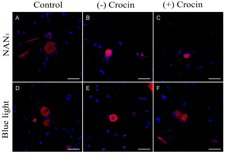
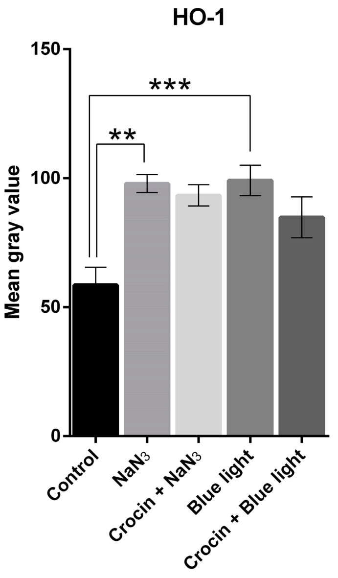
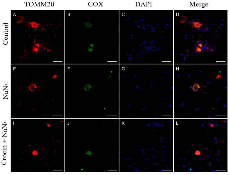
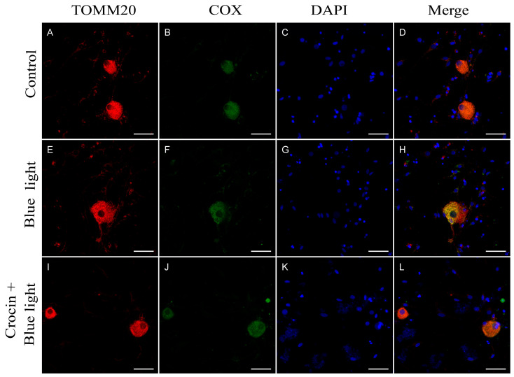
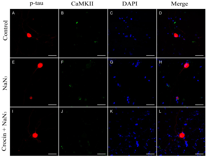
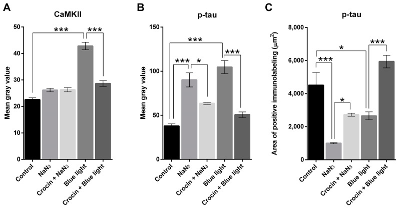
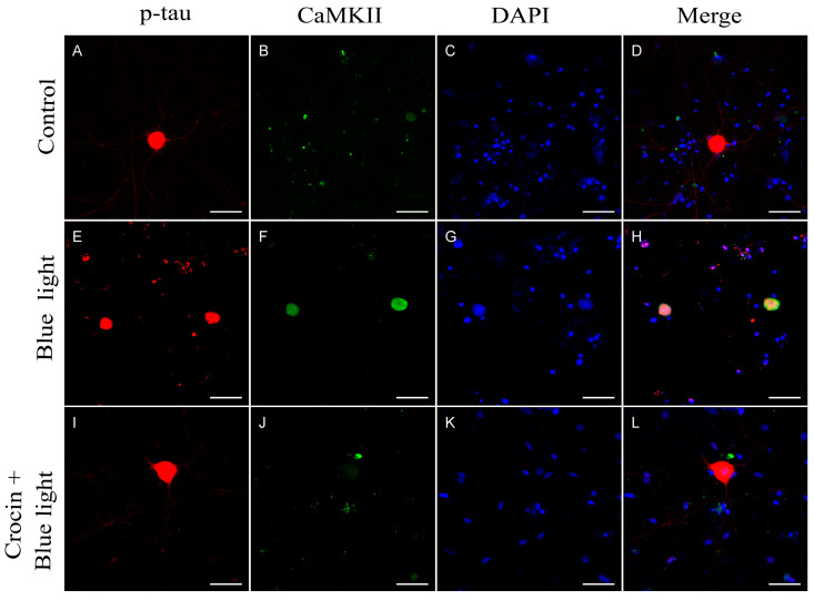
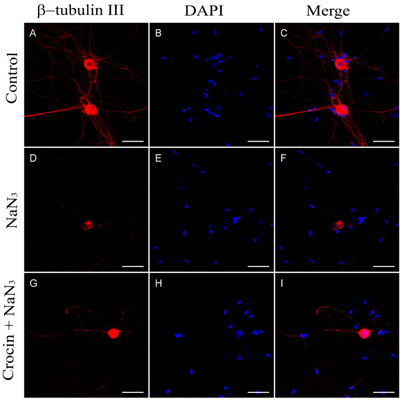
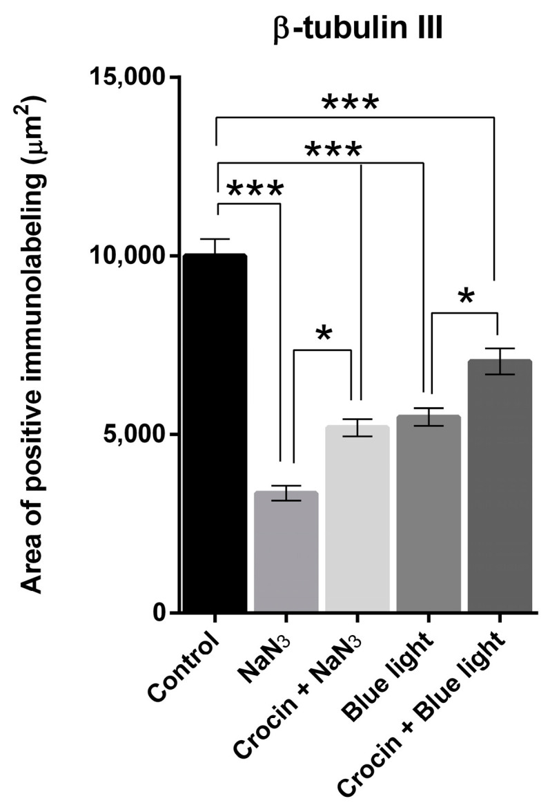
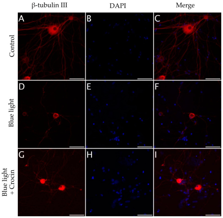
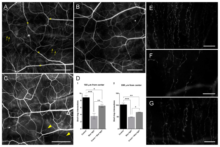
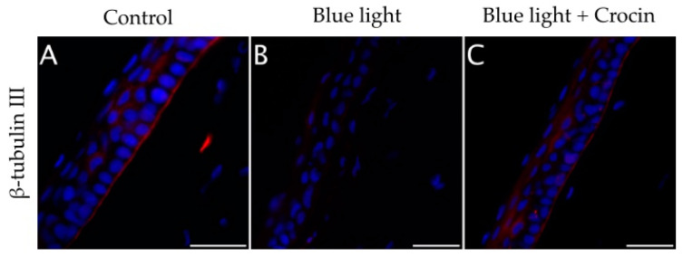
Corneal diseases are a major cause of vision loss, often associated with aging, trauma and disease. Damage to corneal sensory innervation leads to discomfort and pain. Environmental stressors, such as short-wavelength light, can induce oxidative stress that alters mitochondrial function and affects cell and tissue homeostasis, including corneal innervation. Cellular antioxidant mechanisms may attenuate oxidative stress. This study investigates crocin, a derivative of saffron, as a potential antioxidant therapy. In vitro rat trigeminal sensory ganglion neurons were exposed to both sodium azide and blue light overexposure as a model of oxidative damage. Crocin was used as a neuroprotective agent. Mitochondrial and cytoskeletal markers were studied by immunofluorescence analysis to determine oxidative damage and neuroprotection. In vivo corneal innervation degeneration was evaluated in cornea whole mount preparations using Sholl analyses. Blue light exposure induces oxidative stress that affects trigeminal neuron mitochondria and alters sensory axon dynamics in vitro, and it also affects corneal sensory innervation in an in vivo model. Our results show that crocin was effective in preserving mitochondrial function and protecting corneal sensory neurons from oxidative stress. Crocin appears to be a promising candidate for the neuroprotection of corneal innervation.

Keywords: corneal innervation; crocin; neuroprotection; saffron.


Conflict of interest statement

The authors declare no conflicts of interest.

Figures

Figure 1
Figure 1
HO−1 protein detection in undamaged neurons from primary cultures of trigeminal ganglia (A,D) and after exposure to NaN3 (B) and blue light (E). (C,F) show the reduction in fluorescence intensity (HO-1 expression) in cultures treated with crocin for 24 h before exposure to NaN3 (C) and blue light (F). Nuclei were counterstained with DAPI. (Scale bars: 200 µm).
Figure 2
Figure 2
Graph shows the distribution of mean gray values (scaled 0 to 256) corresponding to the expression of HO-1 upon stimulation with NaN3 and blue light compared with crocin-pretreated cultures. A one-way ANOVA with Tukey’s multiple comparisons test was used for parametric data. (Statistical significance is represented by asterisks ** = p < 0.01; *** = p < 0.001).
Figure 3
Figure 3
Immunostaining for TOMM20 and COX. (AD) Neurons from primary cultures of trigeminal ganglia under normal conditions. (EH) Cultures treated with NaN3 for 24 h. Note that in the case of cultures treated with NaN3, there is delocalization of cytochrome C and the mitochondrial marker TOMM20 (H). (IL) Cultures pretreated with crocin before NaN3 damage. Nuclei were counterstained with DAPI. (Scale bars: 200 µm).
Figure 4
Figure 4
Immunostaining for TOMM20 and COX. (AD) Neurons from primary cultures of trigeminal ganglia under normal conditions. (EH) Cultures treated with blue light for 24 h. Note the cleavage of COX in cultures exposed to blue light (H). (IL) Cultures pretreated with crocin before blue light exposure. Nuclei were counterstained with DAPI. (Scale bars: 200 µm).
Figure 5
Figure 5
Expression of p-tau protein and CaMKII in neurons from primary cultures of trigeminal ganglia. (AD) Images from control undamaged cultures showing normal morphology and level of expression of p-tau (A) and CaMKII (B). (EH) Neurons exposed to NaN3 oxidative damage. Note the reduction in neurite number (E) and a slight increase in CaMKII immunofluorescence (F). Cultures pretreated with crocin (IL) showed a significant higher number of neurites (I) and a basal expression of CaMKII (J). Nuclei were counterstained with DAPI. (Scale bars: 200 µm).
Figure 6
Figure 6
Graph (A) shows the distribution of mean gray values (scaled 0 to 256) corresponding to the expression of CaMKII upon stimulation with NaN3 and blue light compared with crocin-pretreated cultures. Graph (B) shows the distribution of mean gray values corresponding to the expression of p-tau protein upon stimulation with NaN3 and blue light compared with crocin-pretreated cultures. Graph (C) shows the relative density (area of positive elements/total area of the selection) corresponding to the expression of p-tau upon stimulation with NaN3 and blue light compared with crocin-pretreated cultures. A one-way ANOVA with Tukey’s multiple comparisons test was used for parametric data. (Statistical significance is represented by asterisks * = p < 0.05; *** = p < 0.001).
Figure 7
Figure 7
Expression of p-tau protein and CaMKII in neurons from primary cultures of trigeminal ganglia. (AD) Images from control undamaged cultures showing normal morphology and level of expression of p-tau (A) and CaMKII (B). (EH) Neurons exposed to blue light damage. Note the reduction in neurite number (E) and a sharp increase in CaMKII immunofluorescence (F). Cultures pretreated with crocin (IL) showed a significant higher number of neurites (I) and a reduced expression of CaMKII (J). Nuclei were counterstained with DAPI. (Scale bars: 200 µm).
Figure 8
Figure 8
(AC) Immunodetection of β-tubulin III in neurons from primary cultures of trigeminal ganglia under normal culture conditions. The addition of NaN3 to the culture medium induced abnormalities and a reduction in neurite density (DF). Pretreatment with crocin for 24 h exerted a partial protective effect on the density of neuronal projections (GI). Nuclei were counterstained with DAPI. (Scale bars: 200 µm).
Figure 9
Figure 9
Graph shows the relative density (area of positive elements/total area of the selection) corresponding to the expression of β-tubulin III upon stimulation with NaN3 and blue light compared with crocin-pretreated cultures. A one-way ANOVA with Tukey’s multiple comparisons test was used for parametric data. (Statistical significance is represented by asterisks * = p < 0.05; *** = p < 0.001).
Figure 10
Figure 10
(AC) Immunodetection of β-tubulin III in neurons from primary cultures of trigeminal ganglia under normal culture conditions. The exposure to blue light induced a reduction in neurite density (DF). Pretreatment with crocin for 24 h exerted a partial protective effect on the density of neuronal projections (GI). Nuclei were counterstained with DAPI. (Scale bars: 200 µm).
Figure 11
Figure 11
Whole mount preparations of rat corneas showing the corneal innervation of a control undamaged animal (A), a cornea exposed to blue light for 4 h over 10 days (B) and a cornea pretreated with topical 0.1 mM crocin over 3 days previous to blue light exposure (C). Arrows in (A) indicate sub-basal nerve fibers. Asterisks in (A) indicate stromal nerve bundles. Note the significantly higher density of sub-basal nerve fibers in (C) compared with (B) (see also (D)). Arrowheads in (C) indicate regenerative milieu at the tip of sub-basal nerve fibers. β-tubulin III immunofluorescence signal was artificially colored to gray in order to increase the visualization of the small sub-basal fibers. Dotted circles indicate the 600 µm Sholl ring. (D) Graphs showing the frequency of intersections of sub-basal nerve fibers with Sholl rings at 100 and 600 µm from the center. A one-way ANOVA with Tukey’s multiple comparisons test was used for parametric data. (E) Detail of sub-basal nerve fibers from image (A) (control undamaged) at the level of the 600 µm Sholl ring. (F) Detail of sub-basal nerve fibers from image (B) (exposed to blue light) at the level of the 600 µm Sholl ring. (G) Detail of sub-basal nerve fibers from image (C) (pretreated with crocin before blue light exposure) at the level of the 600 µm Sholl ring. Note the difference in fiber density between groups. (Scale bars in (AC): 200 µm; scale bars in (EG): 50 µm; statistical significance is represented by asterisks in (D): * = p < 0.05; ** = p < 0.01; *** = p < 0.001).
Figure 12
Figure 12
(A) Cross-sections of the cornea of an undamaged control rat showing a normal distribution of β-tubulin III positive sub-basal nerve plexus at the level of the basement membrane of the corneal epithelium. (B) Corneas exposed to blue light showed a loss of β-tubulin III immunolabeling at the basal epithelium, indicating nerve degeneration. (C) Corneas treated with crocin before blue light insult showed a visible β-tubulin III positive sub-basal plexus. Nuclei were counterstained with DAPI. (Scale bars: 50 µm).

Similar articles

References

    1. Stapleton F., Alves M., Bunya V.Y., Jalbert I., Lekhanont K., Malet F., Na K.S., Schaumberg D., Uchino M., Vehof J., et al. TFOS DEWS II Epidemiology Report. Ocul. Surf. 2017;15:334–365. doi: 10.1016/j.jtos.2017.05.003. - DOI - PubMed
    1. Marfurt C.F., Cox J., Deek S., Dvorscak L. Anatomy of the Human Corneal Innervation. Exp. Eye Res. 2010;90:478–492. doi: 10.1016/j.exer.2009.12.010. - DOI - PubMed
    1. Müller L.J., Marfurt C.F., Kruse F., Tervo T.M.T. Corneal Nerves: Structure, Contents and Function. Exp. Eye Res. 2003;76:521–542. doi: 10.1016/S0014-4835(03)00050-2. - DOI - PubMed
    1. Gonzalez-Coto A.F., Alonso-Ron C., Alcalde I., Gallar J., Meana Á., Merayo-Lloves J., Belmonte C. Expression of Cholecystokinin, Gastrin, and Their Receptors in the Mouse Cornea. Investig. Ophthalmol. Vis. Sci. 2014;55:1965–1975. doi: 10.1167/iovs.13-12068. - DOI - PubMed
    1. Alcalde I., Íñigo-Portugués A., González-González O., Almaraz L., Artime E., Morenilla-Palao C., Gallar J., Viana F., Merayo-Lloves J., Belmonte C. Morphological and Functional Changes in TRPM8-Expressing Corneal Cold Thermoreceptor Neurons during Aging and Their Impact on Tearing in Mice. J. Comp. Neurol. 2018;526:1859–1874. doi: 10.1002/cne.24454. - DOI - PubMed