Review
. 2023 Dec 31;16(1):75.
doi: 10.3390/v16010075.

The Repurposing of Cellular Proteins during Enterovirus A71 Infection

Affiliations
Review

The Repurposing of Cellular Proteins during Enterovirus A71 Infection

Sudeshi M Abedeera et al. Viruses. .

Abstract

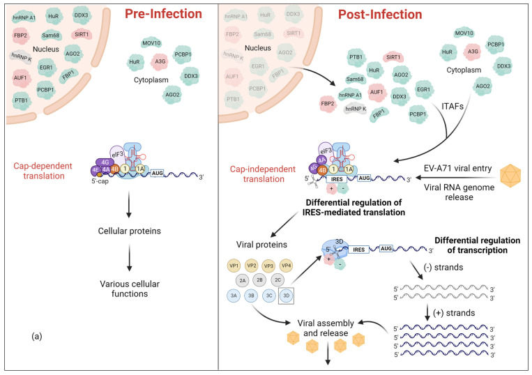
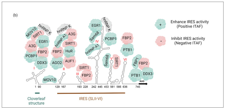
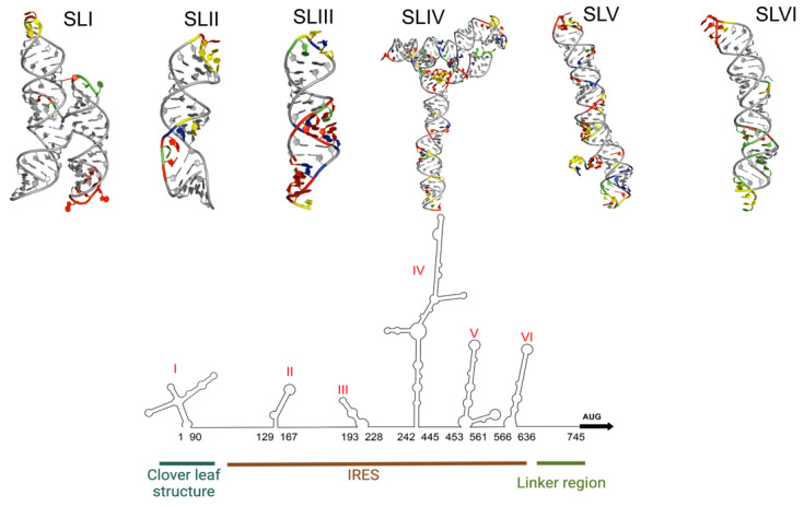
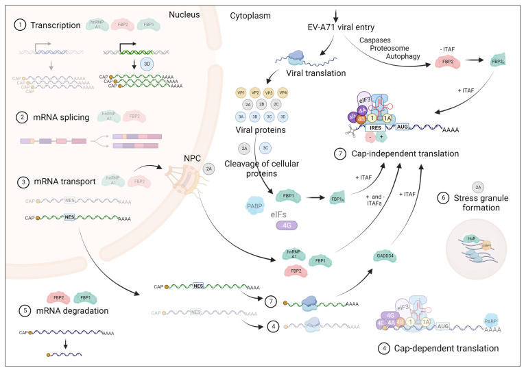
Viruses pose a great threat to people's lives. Enterovirus A71 (EV-A71) infects children and infants all over the world with no FDA-approved treatment to date. Understanding the basic mechanisms of viral processes aids in selecting more efficient drug targets and designing more effective antivirals to thwart this virus. The 5'-untranslated region (5'-UTR) of the viral RNA genome is composed of a cloverleaf structure and an internal ribosome entry site (IRES). Cellular proteins that bind to the cloverleaf structure regulate viral RNA synthesis, while those that bind to the IRES also known as IRES trans-acting factors (ITAFs) regulate viral translation. In this review, we survey the cellular proteins currently known to bind the 5'-UTR and influence viral gene expression with emphasis on comparing proteins' functions and localizations pre- and post-(EV-A71) infection. A comprehensive understanding of how the host cell's machinery is hijacked and reprogrammed by the virus to facilitate its replication is crucial for developing effective antivirals.

Keywords: 5′-UTR; IRES; ITAF; enterovirus A71 (EV-A71).


Conflict of interest statement

The authors declare no conflicts of interest.

Figures

Figure 1
Figure 1
Reprogramming of the cellular environment upon EV-A71 infection. Following viral infection, several host RNA binding proteins, collectively referred to as ITAFs, change their sub-cellular localization. (a) These cellular ITAFs are primarily localized in the nucleus or cytoplasm while some of them can be shuttled between nucleus and cytoplasm based on their cellular functions. Regardless, upon EV-A71 infection, all ITAFs will be relocalized into the cytoplasm to regulate IRES-mediated translation. (b) The 5′-UTR of EV-A71 genome consists of the cloverleaf structure (SLI) and the IRES (SLII-VI). Cellular ITAFs bind to specific region(s) within IRES to either positively or negatively regulate IRES-mediated translation. Positive and negative ITAFs are indicated in green and red, respectively. The ITAFs, of which the effect on translation is not discovered yet, are indicated in grey. Some ITAFs can also bind the SLI region to regulate viral replication.
Figure 1
Figure 1
Reprogramming of the cellular environment upon EV-A71 infection. Following viral infection, several host RNA binding proteins, collectively referred to as ITAFs, change their sub-cellular localization. (a) These cellular ITAFs are primarily localized in the nucleus or cytoplasm while some of them can be shuttled between nucleus and cytoplasm based on their cellular functions. Regardless, upon EV-A71 infection, all ITAFs will be relocalized into the cytoplasm to regulate IRES-mediated translation. (b) The 5′-UTR of EV-A71 genome consists of the cloverleaf structure (SLI) and the IRES (SLII-VI). Cellular ITAFs bind to specific region(s) within IRES to either positively or negatively regulate IRES-mediated translation. Positive and negative ITAFs are indicated in green and red, respectively. The ITAFs, of which the effect on translation is not discovered yet, are indicated in grey. Some ITAFs can also bind the SLI region to regulate viral replication.
Figure 2
Figure 2
The EV-A71 5′-UTR contains phylogenetically conserved stem-loops (SLs) that adopt well-defined 3D structures. Depicted is a predicted secondary structure of the 5′-UTR of EV-A71, and 3D models of each individual stem-loop. The four nucleotides: adenosine (red), uridine (green), guanosine (blue), cytidine (yellow) are color coded in the regions that are predicted to be single stranded and most likely to interact with listed ITAFs. The base-paired nucleotides are shown in grey. 3D structures of SL I and II are published PDB structures while SLs III–VI were produced using the FAFFAR module of the ROSETTA software suit. The lowest energy structure for each stem-loop has been selected from a pool of 10,000 structures. The structures of SLI (8DP3) and SLII (5V17) were obtained from the PDB. Note, the structure of SLI of EV-A71 has not been published yet. Shown here is the SLI of the coxsackievirus B3 (CVB3) that was selected due to the high secondary structural homology between SLI regions of EV-A71 and CVB3.
Figure 3
Figure 3
Cellular ITAFs bind with specificity to short and degenerate sequence motifs. Depicted are published PDB structures of cellular ITAFs bound to RNA. These structures have been experimentally determined by both X-ray and nuclear magnetic resonance (NMR) spectroscopy. RNA has been color coded: adenosine (red), uridine (green), guanosine (blue), cytidine (yellow). ITAFs that bind to double-stranded RNA have the RNA depicted as a cartoon structure. The identity and PDB ids of the depicted ITAFs are as follows: (a) Argonaute 2 (5KI6), (b) hnRNP F (2KFY), (c) Far -Upstream Binding Protein 2 (4B8T), (d) hnRNP A1 (4YOE), (e) Polypyrimidine tract-binding protein 1 (2N3O), (f) Human Antigen R (6G2K), (g) DEAD-Box Protein 3 (6O5F), (h) Staufen homolog 1 (6HTU). Note, the crystal structure of the hnRNP A1-RNA complex (4YOE) was solved with a 5′-AGU-3′ sequence bound to only its RRM1 domain, confirming that a single-strand AG dinucleotide is the minimal specificity motif.
Figure 4
Figure 4
EV-A71 infection perturbs normal RNA metabolism, which leads to a shift in cellular homeostasis. Shown in steps 1–7 are some of the main cellular mechanisms that occur in the host cell. The mechanisms that are illustrated as faded are the ones that are affected by EV-A71 infection and are inhibited specifically due to the relocalization of cellular proteins to be used as ITAFs in EV-A71 IRES-mediated (cap-independent) translation. A few of the ITAFs, hnRNPA1, FBP1, FBP2, GADD34, and HuR, are used to represent the whole repertoire of ITAFs to help explain different viral mechanisms adapted by EV-A71 to facilitate its IRES-mediated translation. Positive ITAFs are colored in green while negative ITAFs are colored in red. These ITAFs are shown at the cellular functions that they regulate prior to EV-A71 infection. Note, a cleaved product of eIF4G is still involved in translation initiation, even though it is shown as faded due to cleavage by viral protease 3Cpro.

References

    1. Nayak G., Bhuyan S.K., Bhuyan R., Sahu A., Kar D., Kuanar A. Global Emergence of Enterovirus 71: A Systematic Review. Beni-Suef Univ. J. Basic Appl. Sci. 2022;11:78. doi: 10.1186/s43088-022-00258-4. - DOI - PMC - PubMed
    1. Hu Y.-L., Chen C.-M., Wang E.-T., Kuo H.-W., Shih W.-L., Fang C.-T., Liu D.-P., Chang L.-Y. The Secular Trend of Enterovirus A71 after the Implementation of Preventive Measures in Taiwan. BMC Public Health. 2022;22:1483. doi: 10.1186/s12889-022-13916-0. - DOI - PMC - PubMed
    1. Enterovirus 71. [(accessed on 8 November 2023)]. Available online: https://www.who.int/teams/health-product-policy-and-standards/standards-....
    1. Huang P.-N., Shih S.-R. Update on Enterovirus 71 Infection. Curr. Opin. Virol. 2014;5:98–104. doi: 10.1016/j.coviro.2014.03.007. - DOI - PubMed
    1. Ibba R., Carta A., Madeddu S., Caria P., Serreli G., Piras S., Sestito S., Loddo R., Sanna G. Inhibition of Enterovirus A71 by a Novel 2-Phenyl-Benzimidazole Derivative. Viruses. 2021;13:58. doi: 10.3390/v13010058. - DOI - PMC - PubMed

LinkOut - more resources