. 2024 Jan 5;16(1):87.
doi: 10.3390/v16010087.

Safety and Efficacy upon Infection in Sheep with Rift Valley Fever Virus ZH548-rA2, a Triple Mutant Rescued Virus

Affiliations

Safety and Efficacy upon Infection in Sheep with Rift Valley Fever Virus ZH548-rA2, a Triple Mutant Rescued Virus

Sandra Moreno et al. Viruses. .

Abstract

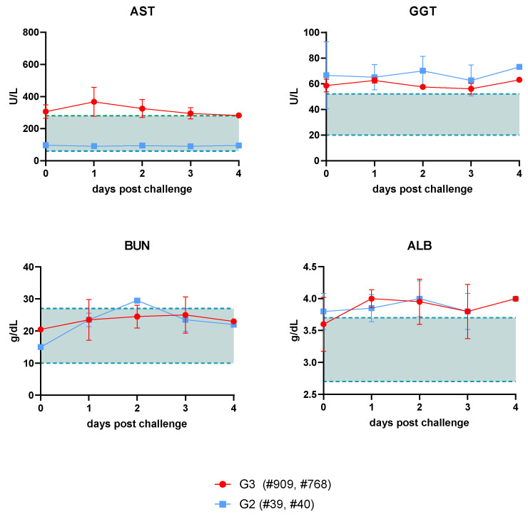
The introduction of three single nucleotide mutations into the genome of the virulent RVFV ZH548 strain allows for the rescue of a fully attenuated virus in mice (ZH548-rA2). These mutations are located in the viral genes encoding the RdRp and the non-structural protein NSs. This paper shows the results obtained after the subcutaneous inoculation of ZH548-rA2 in adult sheep and the subsequent challenge with the parental virus (ZH548-rC1). Inoculation with the ZH548-rA2 virus caused no detectable clinical or pathological effect in sheep, whereas inoculation of the parental rC1 virus caused lesions compatible with viral infection characterised by the presence of scattered hepatic necrosis. Viral infection was confirmed via immunohistochemistry, with hepatocytes within the necrotic foci appearing as the main cells immunolabelled against viral antigen. Furthermore, the inoculation of sheep with the rA2 virus prevented the liver damage expected after rC1 virus inoculation, suggesting a protective efficacy in sheep which correlated with the induction of both humoral and cell-mediated immune responses.

Keywords: Rift Valley fever virus; attenuating mutations; sheep pathology; vaccine efficacy.


Conflict of interest statement

The authors declare no conflict of interests. The funders had no role in the design of the study; in the collection, analyses, or interpretation of data; in the writing of the manuscript; or in the decision to publish the results.

Figures

Figure 1
Figure 1
Experimental outline of the small-scale sheep trial described in this work. Three groups were defined for different aims. A first group (G1) was established for testing immunogenicity and pathology after rA2 virus inoculation. A second group (G2) was established to test efficacy of rA2 as a vaccine after rC1 challenge. The pathology of rC1 was tested in G3. Individual sheep numbers are indicated. Arrowheads indicate blood sample retrieval before (beige) or after challenge (red). Numbers below indicate days after immunisation or challenge. Blood samples were collected in EDTA tubes (for whole blood or plasma-based analysis) or in clotting tubes for serum retrieval.
Figure 2
Figure 2
Daily mean rectal temperatures in rZH548-inoculated sheep. The plots represent mean and standard deviations. (A) Temperature records of the four sheep inoculated (vaccinated) with rA2 virus (G1 and G2) on similar days before and after inoculation (shown up to 7 dpi, as both sheep #73 and #83 were necropsied on this date). (B) Sheep temperatures upon rC1 challenge in both rA2-vaccinated (G2) and non-vaccinated (G3) sheep (for sheep #39 and #40, the −5 to −1 days correspond to 16 to 20 days post vaccination in graph A). The normal rectal temperature range for sheep is shaded between the dashed lines. Data in graphs available as supplementary material.
Figure 3
Figure 3
Blood chemistry analysis in vaccinated and/or challenged sheep (G2 and G3). Hepatic transaminases and protein degradation levels were measured in serum samples taken at the indicated days post challenge with rC1 virus. AST: aspartate aminotransferase. GGT: gamma-glutamyltransferase. ALB: serum albumin. BUN: serum urea nitrogen. The plots represent mean and standard deviations. Shaded areas denote normal enzyme values as described previously [32]. Data in graphs available as supplementary material.
Figure 4
Figure 4
Macroscopic lesions in sheep liver. Representative images of livers taken during necropsies at days 3 (sheep 768) or 4 (sheep 909) post inoculation with rC1 virus (G3). Note the greyish-white foci, many of them surrounded by a reddish halo, scattered throughout the liver parenchyma, and corresponding to areas of necrosis.
Figure 5
Figure 5
Histopathological and immunohistochemical findings observed in the liver of rC1-inoculated sheep (G3). (A) Detection of viral antigen in liver sections via immunohistochemistry using an in-house rabbit anti-RVFV polyclonal serum (upper panel magnification: 10×; lower panel magnification inset: 40×). Note the presence of immunolabelled hepatocytes within the necrotic foci, which were scattered throughout the liver parenchyma. (B) Haematoxylin–eosin stain. Necrotic foci filled with cellular debris and both degenerated and viable neutrophils, surrounded by mononuclear cells, mainly lymphocytes (magnification: 40×). Note the eosinophilic intranuclear inclusion bodies within the hepatocytes (inset arrows; magnification: 100×). Also, note the parallelism between viral antigen-immunolabelled foci and necrotic foci in haematoxylin–eosin sections.
Figure 6
Figure 6
Macroscopic and histopathological evaluation of the liver of sheep inoculated with rA2 virus used as a vaccine candidate. (A) Representative macroscopic images of the liver of sheep #40 (inoculated with rA2 virus, challenged with rC1 virus and euthanised on day 4 post challenge) and sheep #73 (only inoculated with rA2 virus only and euthanised on day 7 post inoculation) to show the absence of macroscopic lesion. Serial sections of the liver of sheep #40 and #73 stained with haematoxylin–eosin (B) and immunolabelled against viral antigen with an in-house rabbit anti-RVFV polyclonal serum (C). Note the absence of liver lesions or cells marked against viral antigen (magnification: 40×).
Figure 7
Figure 7
Analysis of humoral responses in sheep. (A) Kinetics of RVFV-neutralising antibody induction detected in serum microneutralisation test in rA2-inoculated sheep (G2). The measurement of neutralising antibodies in sheep #73 and #83 (G1) was performed only at day 7 post inoculation (symbols labelled with asterisk). Dotted line shows the predicted titre that correlates with protection of the assay. PC: post challenge (3 dpi). (B) Detection of interferon gamma in plasma samples by specific IFNγ capture sandwich ELISA. Challenge was performed at day 21. The plots represent mean and standard error. Data in graphs available as supplementary material.
Figure 8
Figure 8
Determination of RVFV-specific T-cell responses in sheep. Detection of IFNγ in plasma from rA2-inoculated sheep (G2) by capture ELISA (A) Blood samples taken at 7, 14 or 21 days after rA2 inoculation were re-stimulated with recombinant RVFV Gn or N proteins. A recombinant VP2 protein from BTV-8 was used as a negative stimulus (C-). (B) Peptides derived from the Gn (#19 and #21) or Gc (#226 and #253) protein sequences were used for re-stimulation. The plots represent individual sheep values (symbols) and mean and standard deviations (bars). Data in graphs available as supplementary material.

References

    1. Napp S., Chevalier V., Busquets N., Calistri P., Casal J., Attia M., Elbassal R., Hosni H., Farrag H., Hassan N., et al. Understanding the legal trade of cattle and camels and the derived risk of Rift Valley Fever introduction into and transmission within Egypt. PLoS Negl. Trop. Dis. 2018;12:e0006143. doi: 10.1371/journal.pntd.0006143. - DOI - PMC - PubMed
    1. Gibson S., Linthicum K.J., Turell M.J., Anyamba A. Rift Valley fever virus: Movement of infected humans threatens global public health and agriculture. CABI Rev. 2022;17 doi: 10.1079/cabireviews202217029. - DOI
    1. Grobbelaar A.A., Weyer J., Leman P.A., Kemp A., Paweska J.T., Swanepoel R. Molecular epidemiology of Rift Valley fever virus. Emerg. Infect. Dis. 2011;17:2270–2276. doi: 10.3201/eid1712.111035. - DOI - PMC - PubMed
    1. Freiberg A.N., Sherman M.B., Morais M.C., Holbrook M.R., Watowich S.J. Three-dimensional organization of Rift Valley fever virus revealed by cryoelectron tomography. J. Virol. 2008;82:10341–10348. doi: 10.1128/JVI.01191-08. - DOI - PMC - PubMed
    1. Huiskonen J.T., Överby A.K., Weber F., Grünewald K. Electron cryo-microscopy and single-particle averaging of Rift Valley fever virus: Evidence for GN-GC glycoprotein heterodimers. J. Virol. 2009;83:3762–3769. doi: 10.1128/JVI.02483-08. - DOI - PMC - PubMed

Substances

LinkOut - more resources