Review
. 2024 Jan 7;12(1):121.
doi: 10.3390/microorganisms12010121.

The Evolving Microbiome of Dental Caries

Affiliations
Review

The Evolving Microbiome of Dental Caries

Grace Spatafora et al. Microorganisms. .

Abstract

Dental caries is a significant oral and public health problem worldwide, especially in low-income populations. The risk of dental caries increases with frequent intake of dietary carbohydrates, including sugars, leading to increased acidity and disruption of the symbiotic diverse and complex microbial community of health. Excess acid production leads to a dysbiotic shift in the bacterial biofilm composition, demineralization of tooth structure, and cavities. Highly acidic and acid-tolerant species associated with caries include Streptococcus mutans, Lactobacillus, Actinomyces, Bifidobacterium, and Scardovia species. The differences in microbiotas depend on tooth site, extent of carious lesions, and rate of disease progression. Metagenomics and metatranscriptomics not only reveal the structure and genetic potential of the caries-associated microbiome, but, more importantly, capture the genetic makeup of the metabolically active microbiome in lesion sites. Due to its multifactorial nature, caries has been difficult to prevent. The use of topical fluoride has had a significant impact on reducing caries in clinical settings, but the approach is costly; the results are less sustainable for high-caries-risk individuals, especially children. Developing treatment regimens that specifically target S. mutans and other acidogenic bacteria, such as using nanoparticles, show promise in altering the cariogenic microbiome, thereby combatting the disease.

Keywords: Streptococcus mutans; caries; ecology; microbiology; treatment.


Conflict of interest statement

The authors declare no conflicts of interest.

Figures

Figure 1
Figure 1
The distribution of dental caries by age, race, and ethnicity, and family income in the U.S. population. (A) Prevalence of caries in primary teeth. Compared with NHANES 1988–1994, caries experience slightly decreased for children 2- to 5-year-olds. However, the decrease was not observed for 6- to 8-year-old children between 1988 and 2016. (B) Trend in dental caries. There was a slight decrease in caries experience in the permanent dentition of young children and adolescents between 1988 and 2016. However, there are no significant changes in overall caries status in adults aged 20 to 64 years old. Caries prevalence significantly increased for the elderly aged 64 years and over. (CE) Prevalence of caries in primary and permanent teeth in 2011–2016 by age (C), race and ethnicity (D), and family poverty status, FPL = Federal Poverty Level (E). (FH) Prevalence of untreated caries by age (F), race and ethnicity, NHisp = Non Hispanic (G), and family poverty level (FPL) (H) in the U.S. population.
Figure 2
Figure 2
Major species identified from caries-free sites, enamel caries, and dentin/root caries. Shown are cross sections of the enamel, dentin, and root of a tooth with their associated caries-free and caries-associated microbiota. Caries-free microbiotas colonize the tooth surface as diverse communities that respond to dietary carbohydrate challenge by producing acid, favoring enamel demineralization that is neutralized by microbiome community activity that includes the production of ammonia deiminase and urease activity (remineralization). Enamel caries derives from the acid-induced enrichment of highly acidogenic species and suppression of acid-sensitive species, leading to a reduction in community diversity. Caries in dentin, including root caries at the tooth surface, generally comprises a more diverse microbiota than enamel caries with moderately rather than highly acidogenic species owing to the reduced mineral content of dentin. Caries progression deep in dentin can involve acidic demineralization in addition to increasing proteolytic activity, leading to higher pH values in deep lesions. The sequence of caries progression shown is based on Takahashi and Nyvad [73]. Species based on text references.
Figure 3
Figure 3
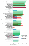
Microbial species and taxa, including viruses, detected from gene expression profiles in coronal and dentin caries. Bacterial samples were taken from coronal caries and caries-free sites and dentin caries from early childhood caries in children with progressing lesions. Functional profiling was performed on purified bacterial RNA using HUMAnN 2.0, version 0.9.9 (HMP Unified Metabolic Analysis Network). Species mapped from gene expression profiles showed the greatest diversity from dentin caries samples. Dentin caries n = 6; coronal caries n = 5; caries n = 4. * difference detection, Chi-square > 0.05. Data from Kressirer et al. [142].
Figure 4
Figure 4
Diversity of oral Lactobacillus species. (A) Composition of Lactobacillus species isolated in the oral cavity of 3- to 5-year-old children (N = 74). (B) Distribution of twenty-one Lactobacillus species identified in children with severe early childhood caries (S-ECC, N = 37) and children without caries (CF, N = 37). The figure shows that the abundance and distribution of the Lactobacillus species were significantly different between the two groups of children [189].
Figure 5
Figure 5
Relative abundance of the 30 most prevalent bacterial genera detected in advanced caries [191]. Bacterial samples were taken from deep occlusal (dentin) caries in permanent molars from 10 individuals. DNA was extracted for 16S rRNA gene (V4 variable region) sequence analysis. Approximately 347,646 partial 16S rRNA gene sequences were obtained. Overall, the Lactobacillus genus accounted for 42.3% of the sequences. The relative abundance per case in five samples ranged from 63% to 96% of the bacterial sequences.
Figure 6
Figure 6
Lactobacillus species detected in vaginal and gingival samples. Oral (gingival) and vaginal samples were taken from 194 pregnant women in the first trimester [196]. Samples were analyzed using DNA gene probes in a checkerboard format with a 105 threshold of species detection. Of the Lactobacillus species assayed, most were detected in both sample sites, suggesting that the vagina could be a source of oral lactobacilli in infants. In contrast, other species typical of subgingival sites, including Porphyromonas and Prevotella species, were detected more frequently in the gingival samples. * Difference detection, Chi-square > 0.05.
Figure 7
Figure 7
Acid production from a glucose solution with added fluoride on in vitro dental biofilms. Effects of a glucose solution with added fluoride on the in vitro pH of dental biofilms. pH dropped in 10-day biofilms after an overlay of glucose or glucose + 10 ppm fluoride [247].
Figure 8
Figure 8
Effects of sodium diamine fluoride (SDF) on S. mutans biofilms cultured on dentin blocks in vitro. Shown are scanning electron micrographs (SEM, left panels) of the biofilm topography and the images from confocal laser scanning microscopy (CSLM, right panels) of the S. mutans biofilm on dentin caries lesions treated with (a) 38% SDF solution plus 5% NaF varnish; (b) 38% SDF solution alone; (c) 5% NaF; and (d) a water-treatment control. The green color represents live bacterial cells; the red color represents dead bacterial cells (magnification ×1000). The green intensity represents the amount of biofilm formed in the carious lesions. Modified based on Yu et al. 2018 [279].

References

    1. Milgrom P. Tooth decay in our poorest children: What can we do? J. Indiana Dent. Assoc. 2000;79:24–26. - PubMed
    1. Pitts N.B., Zero D.T., Marsh P.D., Ekstrand K., Weintraub J.A., Ramos-Gomez F., Tagami J., Twetman S., Tsakos G., Ismail A. Dental caries. Nat. Rev. Dis. Primers. 2017;3:17030. doi: 10.1038/nrdp.2017.30. - DOI - PubMed
    1. Meier T., Deumelandt P., Christen O., Stangl G.I., Riedel K., Langer M. Global burden of sugar-related dental diseases in 168 countries and corresponding health care costs. J. Dent. Res. 2017;96:845–854. doi: 10.1177/0022034517708315. - DOI - PubMed
    1. GBD 2017 Oral Disorders Collaborators. Bernabe E., Marcenes W., Hernandez C.R., Bailey J., Abreu L.G., Alipour V., Amini S., Arabloo J., Arefi Z., et al. Global, regional, and national levels and trends in burden of oral conditions from 1990 to 2017: A systematic analysis for the global burden of disease 2017 study. J. Dent. Res. 2020;99:362–373. doi: 10.1177/0022034520908533. - DOI - PMC - PubMed
    1. Agency for Healthcare Research and Quality . MEPS HC-188B: 2016 Dental Visits. Agency for Healthcare Research and Quality; Rockville, MD, USA: 2018.

LinkOut - more resources