. 2024 Feb 28;9(2):e0060923.
doi: 10.1128/msphere.00609-23. Epub 2024 Jan 23.

Structural determination of Rickettsia lipid A without chemical extraction confirms shorter acyl chains in later-evolving spotted fever group pathogens

Affiliations

Structural determination of Rickettsia lipid A without chemical extraction confirms shorter acyl chains in later-evolving spotted fever group pathogens

Hyojik Yang et al. mSphere. .

Abstract

Rickettsiae are Gram-negative obligate intracellular parasites of numerous eukaryotes. Human pathogens of the transitional group (TRG), typhus group (TG), and spotted fever group (SFG) rickettsiae infect blood-feeding arthropods, have dissimilar clinical manifestations, and possess unique genomic and morphological attributes. Lacking glycolysis, rickettsiae pilfer numerous metabolites from the host cytosol to synthesize peptidoglycan and lipopolysaccharide (LPS). For LPS, O-antigen immunogenicity varies between SFG and TG pathogens; however, lipid A proinflammatory potential is unknown. We previously demonstrated that Rickettsia akari (TRG), Rickettsia typhi (TG), and Rickettsia montanensis (SFG) produce lipid A with long 2' secondary acyl chains (C16 or C18) compared to short 2' secondary acyl chains (C12) in Rickettsia rickettsii (SFG) lipid A. To further probe this structural heterogeneity and estimate a time point when shorter 2' secondary acyl chains originated, we generated lipid A structures for two additional SFG rickettsiae (Rickettsia rhipicephali and Rickettsia parkeri) utilizing fast lipid analysis technique adopted for use with tandem mass spectrometry (FLATn). FLATn allowed analysis of lipid A structure directly from host cell-purified bacteria, providing a substantial improvement over lipid A chemical extraction. FLATn-derived structures indicate SFG rickettsiae diverging after R. rhipicephali evolved shorter 2' secondary acyl chains. While 2' secondary acyl chain lengths do not distinguish Rickettsia pathogens from non-pathogens, in silico analyses of Rickettsia LpxL late acyltransferases revealed discrete active sites and hydrocarbon rulers for long versus short 2' secondary acyl chain addition. Our collective data warrant determining Rickettsia lipid A inflammatory potential and how structural heterogeneity impacts lipid A-host receptor interactions.IMPORTANCEDeforestation, urbanization, and homelessness lead to spikes in Rickettsioses. Vector-borne human pathogens of transitional group (TRG), typhus group (TG), and spotted fever group (SFG) rickettsiae differ by clinical manifestations, immunopathology, genome composition, and morphology. We previously showed that lipid A (or endotoxin), the membrane anchor of Gram-negative bacterial lipopolysaccharide (LPS), structurally differs in Rickettsia rickettsii (later-evolving SFG) relative to Rickettsia montanensis (basal SFG), Rickettsia typhi (TG), and Rickettsia akari (TRG). As lipid A structure influences recognition potential in vertebrate LPS sensors, further assessment of Rickettsia lipid A structural heterogeneity is needed. Here, we sidestepped the difficulty of ex vivo lipid A chemical extraction by utilizing fast lipid analysis technique adopted for use with tandem mass spectrometry, a new procedure for generating lipid A structures directly from host cell-purified bacteria. These data confirm that later-evolving SFG pathogens synthesize structurally distinct lipid A. Our findings impact interpreting immune responses to different Rickettsia pathogens and utilizing lipid A adjuvant or anti-inflammatory properties in vaccinology.

Keywords: FLATn; Rickettsia; Rocky Mountain spotted fever; evolution; lipid A; lipopolysaccharide; pathogenesis; rickettsiosis; spotted fever group.


Conflict of interest statement

The authors declare no conflict of interest.

Figures
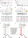
Fig 1
Fig 1
Rickettsia lipid A structures determined by FLATn confirm variable acyl chain lengths for different rickettsiae. (A and B) FLAT-MS spectra from (A) R. rhipicephali partially purified from host cells using either a bead (B) or sucrose gradient (S) purification strategy, and from (B) R. parkeri partially purified from host cells using sucrose gradient purification strategy. Briefly, Vero76 cells were grown to confluence in T-25 flasks with cells infected at an MOI of 10. At 3 days post-infection, host cells were recovered and lysed with 3 mm beads, host debris was removed via low-speed centrifugation (5,000 rpm), and Rickettsia was collected by high-speed centrifugation (8,000 rpm). Purification via sucrose gradient followed our prior protocol (19). Bacterial pellets were then analyzed via FLATn. Asterisks indicate the expected size for Rickettsia lipid A with C16 (~1,936.37 m/z) or C12 (1,880.31 m/z) 2′ secondary acyl chains based on our prior report (19). (C and D) Derivatization of a single ion for the (C) R. rhipicephali sample (1,936.37 m/z) and the (D) R. parkeri sample illustrating five and six, respectively, major fragmentation products. These products are named in the tables with theoretical and experimental sizes shown, with error calculation illustrating robust interpretation. They are also color-coded to facilitate the interpretation of the spectra above and predicted structures. (E and F) FLATn-derived structure predictions for lipid A of (E) R. rhipicephali, which is similar to previously determined R. akari, R. typhi, and R. montanensis structures, and (F) R. parkeri, which is similar to previously determined structures for R. rickettsii strains Shelia Smith and Iowa. Sites yielding fragmentation products are yellow, with corresponding nomenclature described in the tables in panels C and D. The inset describes the conserved and variable lipid A acylation of Rickettsia lipid A, with colored symbols mapped on structures (see Fig. S1 for more details).
Fig 2
Fig 2
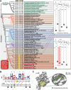
The evolution of variable acyl chain lengths in Rickettsia lipid A. (A) Superimposition of determined lipid A structures and late acyltransferase LpxL characteristics over an estimated Rickettsia phylogeny. Tree is redrawn from a recent study (7). BG, Bellii group and TIG, Tamurae/Ixodes group. Taxon names in gray boxes were determined to synthesize lipid A with palmitate or stearate (C16 or C18) added to the primary 2′ acyl chain; taxon names in red boxes were determined to synthesize lipid A with palmitate or stearate (C16 or C18) added to the primary 2′ acyl chain (see Fig. S1 for more details). P, human pathogen; NP, non-pathogen. Red shading indicates the predicted time point in SFG rickettsiae evolution where a switch from palmitate/stearate to laurate on lipid A 2′ hydroxypalmitate could have occurred based on our structural determinations and shared features of LpxL proteins. Circles 1–5 depict the only variable positions from an alignment of LpxL proteins (panel B). Yellow highlighting indicates a conserved Ile in variable position 3 for the clade-containing species adding laurate on lipid A 2′ hydroxypalmitate. (B) Sequence logo (26) showing conservation of acyltransferase active site residues (asterisks) and five variable positions from an alignment of 127 non-redundant rickettsial LpxL proteins. Alignment was performed using MUSCLE (default parameters) (27). Complete information for all proteins is provided in Table S1. Amino acid coloring scheme and assignment are as follows: black, hydrophobic; red, negatively charged; green, hydrophilic; purple, aromatic; and blue, positively charged. The five variable positions are shown for taxa in the estimated phylogeny in panel A. (C) Visualization of LpxL variable position 3 within the active site of the lipid A late acyltransferase LpxM (note: LpxM is analogous to Rickettsia LpxJ (28)and acylates 3′ acyl chains but was used since no 2′ secondary acyltransferase structures exist). Gray, surface representation of lipid A acyltransferase LpxM from Acinetobacter baumannii (PDBID: 5KN7) is shown in gray. Yellow circle highlighting and insert depict n-dodecyl-β-D-maltoside ligand in green. The acidic active site position (E-127, orange) has a reduced substrate binding when mutated to Ala. Rickettsia variable position 3 (I-130, blue) is found on the same helix near the catalytic E-127 and may be involved in mediating acyl chain length substrate selectivity. Note: 5KN7-N-130 is mutated to I-130 for this depiction; 5KNK is E-127A mutant structure (not shown). Structures are visualized using Chimera (29).

Update of

Similar articles

Cited by

References

    1. Davison HR, Pilgrim J, Wybouw N, Parker J, Pirro S, Hunter-Barnett S, Campbell PM, Blow F, Darby AC, Hurst GDD, Siozios S. 2022. Genomic diversity across the Rickettsia and ‘candidatus megaira’ genera and proposal of genus status for the torix group. Nat Commun 13:2630. doi:10.1038/s41467-022-30385-6 - DOI - PMC - PubMed
    1. Pilgrim J, Thongprem P, Davison HR, Siozios S, Baylis M, Zakharov EV, Ratnasingham S, deWaard JR, Macadam CR, Smith MA, Hurst GDD. 2021. Torix Rickettsia are widespread in arthropods and reflect a neglected symbiosis. Gigascience 10:giab021. doi:10.1093/gigascience/giab021 - DOI - PMC - PubMed
    1. Driscoll TP, Verhoeve VI, Guillotte ML, Lehman SS, Rennoll SA, Beier-Sexton M, Rahman MS, Azad AF, Gillespie JJ, Rikihisa Y. 2017. Wholly Rickettsia ! reconstructed metabolic profile of the quintessential bacterial parasite of eukaryotic cells. mBio 8:e00859-17. doi:10.1128/mBio.00859-17 - DOI - PMC - PubMed
    1. Atwal S, Chuenklin S, Bonder EM, Flores J, Gillespie JJ, Driscoll TP, Salje J. 2021. Discovery of a diverse set of bacteria that build their cell walls without the canonical peptidoglycan polymerase apbp. mBio 12:e0134221. doi:10.1128/mBio.01342-21 - DOI - PMC - PubMed
    1. Figueroa-Cuilan WM, Irazoki O, Feeley M, Smith E, Nguyen T, Cava F, Goley ED. 2023. Quantitative analysis of morphogenesis and growth dynamics in an obligate intracellular bacterium. Mol Biol Cell 34:ar69. doi:10.1091/mbc.E23-01-0023 - DOI - PMC - PubMed

Supplementary concepts