. 2024 Feb 28;9(2):e0052623.
doi: 10.1128/msphere.00526-23. Epub 2024 Jan 23.

Human B cells and dendritic cells are susceptible and permissive to enterovirus D68 infection

Affiliations

Human B cells and dendritic cells are susceptible and permissive to enterovirus D68 infection

Brigitta M Laksono et al. mSphere. .

Abstract

Enterovirus D68 (EV-D68) is predominantly associated with mild respiratory infections, but can also cause severe respiratory disease and extra-respiratory complications, including acute flaccid myelitis. Systemic dissemination of EV-D68 is crucial for the development of extra-respiratory diseases, but it is currently unclear how EV-D68 spreads systemically (viremia). We hypothesize that immune cells contribute to the systemic dissemination of EV-D68, as this is a mechanism commonly used by other enteroviruses. Therefore, we investigated the susceptibility and permissiveness of human primary immune cells for different EV-D68 isolates. In human peripheral blood mononuclear cells inoculated with EV-D68, only B cells were susceptible but virus replication was limited. However, in B cell-rich cultures, such as Epstein-Barr virus-transformed B-lymphoblastoid cell line (BLCL) and primary lentivirus-transduced B cells, which better represent lymphoid B cells, were productively infected. Subsequently, we showed that dendritic cells (DCs), particularly immature DCs, are susceptible and permissive for EV-D68 infection and that they can spread EV-D68 to autologous BLCL. Altogether, our findings suggest that immune cells, especially B cells and DCs, could play an important role in the pathogenesis of EV-D68 infection. Infection of these cells may contribute to systemic dissemination of EV-D68, which is an essential step toward the development of extra-respiratory complications.IMPORTANCEEnterovirus D68 (EV-D68) is an emerging respiratory virus that has caused outbreaks worldwide since 2014. EV-D68 infects primarily respiratory epithelial cells resulting in mild respiratory diseases. However, EV-D68 infection is also associated with extra-respiratory complications, including polio-like paralysis. It is unclear how EV-D68 spreads systemically and infects other organs. We hypothesized that immune cells could play a role in the extra-respiratory spread of EV-D68. We showed that EV-D68 can infect and replicate in specific immune cells, that is, B cells and dendritic cells (DCs), and that virus could be transferred from DCs to B cells. Our data reveal a potential role of immune cells in the pathogenesis of EV-D68 infection. Intervention strategies that prevent EV-D68 infection of immune cells will therefore potentially prevent systemic spread of virus and thereby severe extra-respiratory complications.

Keywords: B cells; dendritic cells; enterovirus; enterovirus D68; extra-respiratory infection; immune cells; systemic spread; viral pathogenesis.


Conflict of interest statement

The authors declare no conflict of interest.

Figures

Fig 1
Fig 1
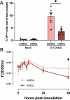
Susceptibility and permissiveness of human PBMC to EV-D68 infection. (A) Percentage of EV-D68 VP1+ immune cell subsets 24 h after inoculation with EV-D68 strains from subclades A (n = 10 donors), B2 (n = 6 donors), and A2 (n = 4 donors). Each symbol represents one donor. Statistical analysis was performed using unpaired t-test. Error bars denoted SEM. (B) Production of infectious virus in EV-D68-inoculated PBMC. PBMC, peripheral blood mononuclear cells; SEM, standard error of mean **P < 0.01.
Fig 2
Fig 2
Susceptibility and permissiveness of B cell-rich models to EV-D68 infection. BLCL and lentivirus-transduced B cells were inoculated with EV-D68 strains from subclades A (n = 7), B2 (n = 6), and A2 (n = 6) as models for EV-D68 infection in B cell-rich environment. (A) Percentage of EV-D68 VP1+ cells at 24 hpi and (B) production of infectious viruses in EV-D68-inoculated BLCL over time, respectively. (C) Percentage of VP1+ cells at 24 hpi and (D) production of infectious viruses in EV-D68-inoculated lentivirus-transduced B-cells. Each symbol in (A) and (C) represents one donor. No statistically significant differences were observed in the percentages of VP1+ cells among the different virus subclades. Statistical analysis was performed using a one-way analysis of variance with multiple comparison test. Error bars denote SEM. BLCL, B-lymphoblastoid cell line; hpi, hours post-inoculation; SEM, standard error of mean.
Fig 3
Fig 3
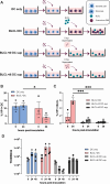
Percentages of α2,3-and α2,6-linked SAs+ and EV-D68 VP1+ BLCL upon neuraminidase treatment. (A) Percentage of BLCL expressed α2,3- (n = 3) and α2,6- (n = 3) linked SAs with and without ANA treatment. (B) Percentage of VP1+ BLCL (n = 3) with and without ANA treatment inoculated with EV-D68 from subclades A, B2, and A2, measured 24 hpi. Statistical analysis was performed with unpaired t-test. Error bars denote SEM. BLCL, B-lymphoblastoid cell line; ANA, Arthrobacter ureafaciens neuraminidase; SAs, sialic acids; hpi, hours post-inoculation; SEM, standard error of mean. *P < 0.05; ****P ≤ 0.0001.
Fig 4
Fig 4
Susceptibility and permissiveness of imDCs and mDCs to EV-D68 infection. (A) Percentage of EV-D68 VP1+ imDCs (n = 5) and mDCs (n = 5) measured at 6 hpi. (B) Production of infectious viruses in EV-D68/A-inoculated imDCs (n = 3) and mDCs (n = 3). Samples for virus titration were collected at 0, 2, 4, 6, 8, 10, 24, and 48 hpi. All statistical analyses in this figure were performed with unpaired t-test. Error bars denote SEM. imDCs, immature dendritic cells; mDCs, mature dendritic cells; hpi, hours post-inoculation; SEM, standard error of mean. *P < 0.05.
Fig 5
Fig 5
Co-culture of EV-D68-inoculated imDCs with their autologous BLCL. (A) Schematic representation of the experimental setup and controls. (B) Percentages of EV-D68 VP1+ imDCs and (C) BLCL in different culture conditions at 6 and 24 hpi. (D) Production of infectious viruses in different culture conditions. Statistical analysis in (B) was performed with unpaired t-test. Statistical analysis in (C) and (D) were performed with a one-way analysis of variance with multiple comparison test and compared to BLCL + t0 DC sup and BLCL + t6 sup, respectively. Error bars denote SEM. BLCL, B-lymphoblastoid cell line; imDCs, immature dendritic cells; hpi, hours post-inoculation; SEM, standard error of mean; BLCL + t0 DC sup, autologous BLCL co-cultured with supernatant collected from EV-D68/A-inoculated imDCs at 0 hpi. BLCL + t6 DC sup, autologous BLCL co-cultured with supernatant collected from EV-D68/A-inoculated imDCs at 6 hpi. *P < 0.05; ***P ≤ 0.001.
Fig 6
Fig 6
Proposed model for systemic dissemination of EV-D68. EV-D68 enters the respiratory tract and initially infects respiratory epithelial cells. The infection will result in the recruitment of immune cells, including imDCs. Subsequently, cell-free EV-D68 can spread to lymphoid tissues by spilling over into the circulatory or lymphatic system. Alternatively, it can infect imDCs, which can transfer the virus to lymphoid tissues. These lymphoid tissues can function as a secondary replication site, where EV-D68 infects mDCs and B cells. From this site, cell-free virus or virus-infected immune cells can enter the blood circulation and spread virus to other tissues. HEV, high endothelial venule; red arrow, blood circulation; purple dotted arrow, potential routes of EV-D68 spread.
Fig 7
Fig 7
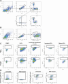
Representative gating strategies for flow cytometry analyses. (A) Gating strategy to determine peripheral blood mononuclear cell subpopulations. B cells were defined as CD3CD19+ cells; CD4+T cells as CD3+CD19CD4+CD8 cells; CD8+T cells as CD3+CD19CD4CD8+ cells; and monocytes as CD3CD19CD14−/+CD16−/+ cells. (B) Gating strategy of EV-D68-inoculated immature dendritic cells (imDCs) co-cultured with autologous B-lymphoblastoid cell line (BLCL). imDCs were defined as CD19 cells; BLCL were defined as CD19+ cells. (C) Gating strategy to determine EV-D68 VP1+ cells. PBMC, peripheral blood mononuclear cells; BLCL, B-lymphoblastoid cell line; DCs, dendritic cells. (D) Gating strategy to determine percentages of α2,3- and α2,6-linked sialic acid+ (SAs+) BLCL. Percentages of SA+ cells were determined at 0 h post-inoculation. ANA, Arthrobacter ureafaciens neuraminidase; BLCL, B-lymphoblastoid cell line.

Similar articles

Cited by

References

    1. Greninger AL, Naccache SN, Messacar K, Clayton A, Yu G, Somasekar S, Federman S, Stryke D, Anderson C, Yagi S, Messenger S, Wadford D, Xia D, Watt JP, Van Haren K, Dominguez SR, Glaser C, Aldrovandi G, Chiu CY. 2015. A novel outbreak enterovirus D68 strain associated with acute flaccid myelitis cases in the USA (2012-14): a retrospective cohort study. Lancet Infect Dis 15:671–682. doi:10.1016/S1473-3099(15)70093-9 - DOI - PMC - PubMed
    1. Messacar K, Abzug MJ, Dominguez SR. 2016. The emergence of enterovirus-D68. Microbiol Spectr 4. doi:10.1128/microbiolspec.EI10-0018-2016 - DOI - PubMed
    1. Messacar K, Schreiner TL, Van Haren K, Yang M, Glaser CA, Tyler KL, Dominguez SR. 2016. Acute flaccid myelitis: a clinical review of US cases 2012-2015. Ann Neurol 80:326–338. doi:10.1002/ana.24730 - DOI - PMC - PubMed
    1. Messacar K, Pretty K, Reno S, Dominguez SR. 2019. Continued biennial circulation of enterovirus D68 in Colorado. J Clin Virol 113:24–26. doi:10.1016/j.jcv.2019.01.008 - DOI - PubMed
    1. Kramer R, Sabatier M, Wirth T, Pichon M, Lina B, Schuffenecker I, Josset L. 2018. Molecular diversity and biennial circulation of enterovirus D68: a systematic screening study in Lyon, France, 2010 to 2016. Euro Surveill 23:1700711. doi:10.2807/1560-7917.ES.2018.23.37.1700711 - DOI - PMC - PubMed

LinkOut - more resources