. 2024 Jan 22;4(1):170-181.
doi: 10.1158/2767-9764.CRC-23-0316.

Lung Fibroblasts Take up Breast Cancer Cell-derived Extracellular Vesicles Partially Through MEK2-dependent Macropinocytosis

Affiliations

Lung Fibroblasts Take up Breast Cancer Cell-derived Extracellular Vesicles Partially Through MEK2-dependent Macropinocytosis

Yuhao Wan et al. Cancer Res Commun. .

Abstract

Extracellular vesicles (EV) have emerged as critical effectors in the cross-talk between cancer and normal cells by transferring intracellular materials between adjacent or distant cells. Previous studies have begun to elucidate how cancer cells, by secreting EVs, adapt normal cells at a metastatic site to facilitate cancer cell metastasis. In this study, we utilized a high-content microscopic screening platform to investigate the mechanisms of EV uptake by primary lung fibroblasts. A selected library containing 90 FDA-approved anticancer drugs was screened for the effect on fibroblast uptake of EVs from MDA-MB-231 breast cancer cells. Among the drugs identified to inhibit EV uptake without exerting significant cytotoxicity, we validated the dose-dependent effect of Trametinib (a MEK1/2 inhibitor) and Copanlisib (a PI3K inhibitor). Trametinib suppressed macropinocytosis in lung fibroblasts and inhibited EV uptake with a higher potency comparing with Copanlisib. Gene knockdown and overexpression studies demonstrated that uptake of MDA-MB-231 EVs by lung fibroblasts required MEK2. These findings provide important insights into the mechanisms underlying lung fibroblast uptake of breast cancer cell-derived EVs, which could play a role in breast cancer metastasis to the lungs and suggest potential therapeutic targets for preventing or treating this deadly disease.

Significance: Through a phenotypic screen, we found that MEK inhibitor Trametinib suppressed EV uptake and macropinocytosis in lung fibroblasts, and that EV uptake is mediated by MEK2 in these cells. Our results suggest that MEK2 inhibition could serve as a strategy to block cancer EV uptake by lung fibroblasts.


Figures

FIGURE 1
FIGURE 1
The high-content microscopic platform for quantitative measurement of EV uptake. A, A schematic graph showing how cellular uptake of fluorescently-labeled EVs was detected and quantified. Briefly, cells were prestained with CFSE (green) and EVs were prestained with DiI (red). Following the incubation, extracellular EVs were washed off and cell nuclei were stained with Hoechst 33342 (blue). During fluorescent image analysis, the CFSE and Hoechst 33342 signals were used to delineate single-cell boundaries and the DiI signals per cell were thereafter quantified. ObjectID, ID for single cells. MFI, mean fluorescent intensity. B, Representative images showing the single-cell delineation process. Human lung fibroblasts were captured at 10 ×. Scale bar, 100 µm. C, Dose-dependent uptake of MDA-MB-231 EVs by human lung fibroblasts. Indicated dosages of EVs were added to cells growing in 100 µL of medium on a 96-well plate for an incubation of 3 hours. Five independent images were taken from each well. The results are presented as the calculated mean value of all MFI in a given well (left; n = 3 independent wells; data shown as mean ± SD) or as the individual MFI of each single cell in that group (right; n > 1,000 cells). In the right panel, the line represents the median of that group and the percentage represents the portion of cells showing an MFI value > 0. Dye-only ctrl, no EVs was added in the EV dye labeling step. ***, P < 0.001; ns, not significant. D, Time-dependent uptake of MDA-MB-231 EVs by human lung fibroblasts. An equal amount of EVs (10 µg/mL) were added to the cells for the indicated periods of incubation. Results are presented as in C. Left: n = 3 independent wells; right: n > 7,000 cells. “1 (on ice)”, cells were incubated with EVs on ice for 1 hour. ***, P < 0.001. E, Representative images from dose-dependent and time-dependent experiments. Images were captured at 10 ×. Green, CFSE; yellow, DiI; blue, Hoechst 33342. Scale bar, 100 µm.
FIGURE 2
FIGURE 2
Human lung fibroblasts take up MDA-MB-231 EVs via dynamin- and caveolae-dependent endocytosis and macropinocytosis. A, Fibroblasts were pretreated with 80 µmol/L Dynasore or an equal volume of DMSO (as a control) for 24 hours and then incubated with 10 µg/mL EVs for 6 hours in the continuous presence of Dynasore or control (n = 3 wells per group; 5 images per well). Data are presented as mean ± SD. ***, P < 0.001. B, Cells were pretreated with 50 µmol/L EIPA, 10 µmol/L CPZ, or 200 µmol/L Genistein for 24 hours and then incubated with 10 µg/mL EVs for 6 hours in the presence of drug (n = 3 wells per group; 5 images per well). Data are presented as mean ± SD. ***, P < 0.001; ns, not significant. C, Representative images of Dynasore-, EIPA-, CPZ-, or Genistein-treated cells and untreated cells. Images were captured at 10 ×. Green, CFSE; yellow, DiI; blue, Hoechst 33342. Scale bar, 100 µm.
FIGURE 3
FIGURE 3
Screening of a targeted library of oncology drugs by high-content microscopy identifies compounds suppressing EV uptake. A, Cells were pretreated with drugs (200 nmol/L final concentration) for 24 hours and then either assayed for viability or incubated with 10 µg/mL EV for 6 hours and assayed for EV uptake. Dynasore (80 µmol/L) was also included as a control treatment. For both MTS assay and EV uptake assay, n = 2 wells per drug were tested. For EV uptake assay, 5 images per well were captured and analyzed. Each dot represents the mean value of two replicates. Drugs resulting in >75% cell viability and <75% EV uptake efficiency were indicated in red. B, Representative images of untreated cells and cells treated with selected drugs. Images were captured at 10 ×. Green, CFSE; yellow, DiI; blue, Hoechst 33342. Scale bar, 100 µm. C, Dose-dependent analysis of the effect of Trametinib and Copanlisib. Experiments were carried out as in A using indicated concentrations of the drug. Data are represented as mean ± SD (n = 3 wells per group; 5 images per well). The best-fit IC50 of each drug is indicated. D, Validation of the effect of Trametinib and Copanlisib by flow cytometry. Experiments were carried out as in A but scaled up to a 6-well plate format. Data are represented as mean ± SD (n = 3 wells). ***, P < 0.001.
FIGURE 4
FIGURE 4
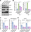
Pathway inhibitors Trametinib and Copanlisib inhibit the uptake and effect of EVs from various breast cancer cells. A, Western blots of lung fibroblasts treated with 200 nmol/L of Trametinib or Copanlisib or with 10 µg/mL EV for 24 hours. B, qRT-PCR–determined mRNA levels of S100A4 and FN1 with or without MDA-MB-231 EV treatment for 48 hours. Trametinib and Copanlisib were added together with EVs when indicated. Data were normalized to GAPDH. C, Lung fibroblasts were pretreated with Trametinib or Copanlisib (200 mmol/L) for 24 hours before Lck-GFP–labeled EVs derived from 4T1 or MDA-MB-468 cells were added and incubated for 6 hours. EV uptake was assessed and compared with DMSO-treated (No drug) control group. D, NIH3T3 fibroblasts were pretreated with Trametinib or Copanlisib before Lck-GFP–labeled EVs derived from 4T1 or MDA-MB-231 cells were added. Data are represented as mean ± SD (n = 3 wells). **, P < 0.01; ***, P < 0.001.
FIGURE 5
FIGURE 5
Trametinib interferes with macropinocytosis in lung fibroblasts while Copanlisib interferes with both macropinocytosis and clathrin-mediated endocytosis. A, Quantification of the cellular uptake of indicated markers with or without Copanlisib or Trametinib treatment. Data are presented as mean ± SD (n = 3 wells per group; 5 images per well). Faint symbols represent all 15 images per group. Dark symbols represent the mean value from each well. **, P < 0.01; ns, not significant. B, Representative images of the cellular uptake of indicated markers. Images were captured at 10 ×. Green, FITC-transferrin, Alexa488-BSA or FITC-dextran; blue, Hoechst 33342. Scale bar, 100 µm.
FIGURE 6
FIGURE 6
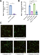
MDA-MB-231 EV uptake by lung fibroblasts requires MEK2 but not MEK1. A, Fibroblasts were transfected with siRNAs against MEK1 or MEK2 (two independent siRNAs used for each gene), or with a control siRNA, at 48 hours before EVs were added for an incubation of 6 hours (n = 3 wells per group). B, Western blots of cells transfected with indicated siRNAs showing the gene knockdown efficiency of MEK1/2. C, Representative images of untreated cells and cells transfected with indicated siRNAs. Images were captured at 10 ×. Green, CFSE; yellow, DiI; blue, Hoechst 33342. Scale bar, 100 µm. D, Fibroblasts were transfected with an overexpression plasmid encoding wild-type (WT) or catalytically inactive (K101A) MEK2, or with the empty vector. Forty-eight hours later, cells were incubated with MDA-MB-231 EVs for 6 hours (n = 3 wells per group). E, Western blots of cells transfected with indicated overexpression plasmids. Data are presented as mean ± SD. *, P < 0.05; **, P < 0.01; ***, P < 0.001; ns, not significant.

Similar articles

Cited by

References

    1. Becker A, Thakur BK, Weiss JM, Kim HS, Peinado H, Lyden D. Extracellular vesicles in cancer: cell-to-cell mediators of metastasis. Cancer Cell 2016;30:836–48. - PMC - PubMed
    1. Wang SE. Extracellular vesicles and metastasis. Cold Spring Harb Perspect Med 2020;10:a037275. - PMC - PubMed
    1. Mitchell PS, Parkin RK, Kroh EM, Fritz BR, Wyman SK, Pogosova-Agadjanyan EL, et al. . Circulating microRNAs as stable blood-based markers for cancer detection. Proc Natl Acad Sci U S A 2008;105:10513–8. - PMC - PubMed
    1. Wu X, Somlo G, Yu Y, Palomares MR, Li AX, Zhou W, et al. . De novo sequencing of circulating miRNAs identifies novel markers predicting clinical outcome of locally advanced breast cancer. J Transl Med 2012;10:42. - PMC - PubMed
    1. Wang SE. Extracellular vesicles in cancer therapy. Semin Cancer Biol 2022;86:296–309. - PMC - PubMed

Publication types

Substances