

An in vitro study of micromechanics, cellular proliferation and viability on both decellularized porcine dura grafts and native porcine dura grafts
- PMID: 38259430
- PMCID: PMC10803071
- DOI: 10.1016/j.bea.2023.100108
An in vitro study of micromechanics, cellular proliferation and viability on both decellularized porcine dura grafts and native porcine dura grafts
Abstract
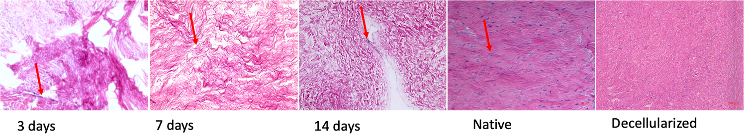
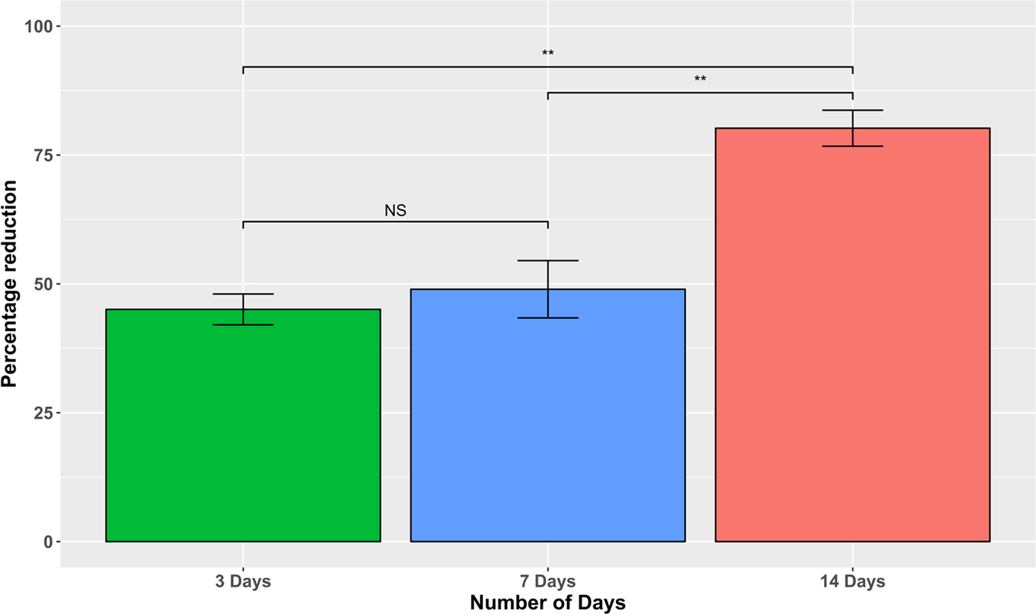
Damage to the dura mater may occur during intracranial or spinal surgeries, which can result in cerebrospinal fluid leakage and other potentially fatal physiological changes. As a result, biological and synthetic derived scaffolds are typically used to repair dura mater post intracranial or spinal surgeries. The extracellular matrix of xenogeneic dura scaffolds has been shown to exhibit increased cell infiltration and regeneration than synthetic dura materials. In this study, we investigated the biocompatibility of native and decellularized porcine dura by seeding rat fibroblast cells onto the constructs. Cell proliferation, cell viability, and the mechanical properties of these dural grafts were evaluated post-re-seeding on days 3,7 and 14. Live-dead staining and resazurin salts were used to quantify cell viability and cell proliferation, respectively. Micro indentation was conducted to quantify the mechanical integrity of the native and acellular dura graft. The findings indicate that the acellular porcine dura graft creates a beneficial setting for infiltrating rat fibroblast cells. Cell viability, proliferation, and micro indentation results on the acellular grafts are comparable with the native control porcine dura tissue. In conclusion, the porcine scaffold material showed increased cell viability at each time point evaluated. The sustained mechanical response and favorable viability of the cells on the decellularized grafts provide promising insight into the potential use of porcine dura in clinical cranial dura mater graft applications.
Keywords: Cranial dura mater; Decellularization; Native dura; Porcine dura.
Conflict of interest statement
Declaration of Competing Interest The authors declare that they have no known competing financial interests or personal relationships that could have appeared to influence the work reported in this paper.
Figures


References
-
- Sharma A, Liao J, Williams LN, Structure and mechanics of native and decellularized porcine cranial dura mater, Eng. Regener 4 (2023) 205–213, 10.1016/j.engreg.2023.02.004. - DOI
-
- O’Brien FJ, Biomaterials & scaffolds for tissue engineering, Mater. Today 14 (3) (2011) 88–95, 10.1016/S1369-7021(11)70058-X. - DOI
Grants and funding
LinkOut - more resources
Full Text Sources
Research Materials