. 2024 Jan 8:14:1334800.
doi: 10.3389/fimmu.2023.1334800. eCollection 2023.

Polymeric nanocapsules loaded with poly(I:C) and resiquimod to reprogram tumor-associated macrophages for the treatment of solid tumors

Affiliations

Polymeric nanocapsules loaded with poly(I:C) and resiquimod to reprogram tumor-associated macrophages for the treatment of solid tumors

Clément Anfray et al. Front Immunol. .

Abstract

Background: In the tumor microenvironment (TME), tumor-associated macrophages (TAMs) play a key immunosuppressive role that limits the ability of the immune system to fight cancer. Toll-like receptors (TLRs) ligands, such as poly(I:C) or resiquimod (R848) are able to reprogram TAMs towards M1-like antitumor effector cells. The objective of our work has been to develop and evaluate polymeric nanocapsules (NCs) loaded with poly(I:C)+R848, to improve drug stability and systemic toxicity, and evaluate their targeting and therapeutic activity towards TAMs in the TME of solid tumors.

Methods: NCs were developed by the solvent displacement and layer-by-layer methodologies and characterized by dynamic light scattering and nanoparticle tracking analysis. Hyaluronic acid (HA) was chemically functionalized with mannose for the coating of the NCs to target TAMs. NCs loaded with TLR ligands were evaluated in vitro for toxicity and immunostimulatory activity by Alamar Blue, ELISA and flow cytometry, using primary human monocyte-derived macrophages. For in vivo experiments, the CMT167 lung cancer model and the MN/MCA1 fibrosarcoma model metastasizing to lungs were used; tumor-infiltrating leukocytes were evaluated by flow cytometry and multispectral immunophenotyping.

Results: We have developed polymeric NCs loaded with poly(I:C)+R848. Among a series of 5 lead prototypes, protamine-NCs were selected based on their physicochemical properties (size, charge, stability) and in vitro characterization, showing good biocompatibility on primary macrophages and ability to stimulate their production of T-cell attracting chemokines (CXCL10, CCL5) and to induce M1-like macrophages cytotoxicity towards tumor cells. In mouse tumor models, the intratumoral injection of poly(I:C)+R848-protamine-NCs significantly prevented tumor growth and lung metastasis. In an orthotopic murine lung cancer model, the intravenous administration of poly(I:C)+R848-prot-NCs, coated with an additional layer of HA-mannose to improve TAM-targeting, resulted in good antitumoral efficacy with no apparent systemic toxicity. While no significant alterations were observed in T cell numbers (CD8, CD4 or Treg), TAM-reprogramming in treated mice was confirmed by the relative decrease of interstitial versus alveolar macrophages, having higher CD86 expression but lower CD206 and Arg1 expression in the same cells, in treated mice.

Conclusion: Mannose-HA-protamine-NCs loaded with poly(I:C)+R848 successfully reprogram TAMs in vivo, and reduce tumor progression and metastasis spread in mouse tumors.

Keywords: antitumoral immunotherapy; cancer; immunotoxicology; poly(I:C); polymeric nanocapsules; resiquimod (R848); toll-like receptor (TLR); tumor-associated macrophages (TAMs).


Conflict of interest statement

The authors declare that the research was conducted in the absence of any commercial or financial relationships that could be construed as a potential conflict of interest. The author(s) declared that they were an editorial board member of Frontiers, at the time of submission. This had no impact on the peer review process and the final decision.

Figures

Figure 1
Figure 1
Polymeric nanocapsules: characterization, toxicity and immunomodulatory evaluation in vitro using primary human macrophages. (A) Schematic representation of the nanocapsules (NCs) and their composition. (B) Dose-response toxicity of blank-NCs on HMDMs exposed for 24h (1; 10; 100 µg/mL); N=4. (C) Physicochemical characteristics of R848-loaded-NCs prepared with different outer polymers (n ≥ 3). (D) Dose-response toxicity on HMDMs exposed for 24h to R848-NCs (0.5 µg/mL); N=9. (E) IC50 values of R848-NCs in HMDMs (24h); N=4. (F) Secretion of CXCL10, CCL5 and IL-6 by HMDMs exposed for 24h to R848-NCs (0.5 µg/mL). (G) Cytotoxic activity of HMDMs, after 48h treatment with R848-NCs (0.5 µg/mL), towards human PANC-1 cells. All values represent mean ± SD. Statistical comparison was performed using a two-way ANOVA followed by a Tuckey’s multiple comparison test. Statistically significant differences are represented as *(p<0.05), **(p<0.01), ***(p<0.001) and ****(p<0.0001). NCs, nanocapsules; PolyArg, polyarginine; Chit, chitosan; Prot, protamine; Dex, dextran sulfate; PSA, poly-sialic-acid.
Figure 2
Figure 2
Synthesis and characterization of poly(I:C)+R848-loaded-protamine-nanocapsules. (A) Schematic representation for the addition of poly(I:C) on the surface protamine coating of the R848-NCs, and description of their components. (B) Physicochemical characterization of protamine-NCs loaded with poly(I:C) and/or R848, and coated with HA or HA-mannose (n ≥ 3). (C) Asymmetrical flow field-flow fractionation (AF4) comparison of R848-loaded protamine nanocapsules (R848-prot-NCs) (left) and poly(I:C)+R848-loaded protamine nanocapsules (pIC+R848-prot-NCs) (right). (D) (left gel) Agarose gel retardation assay to evaluate poly(I:C) binding to the R848-prot-NCs. Controls: (1) free poly(I:C) and (2) free poly(I:C) with heparin; (3) pIC+R848-prot-NCs and (4) pIC+R848-prot-NCs with heparin. (right gel) Agarose gel retardation assay to evaluate the release and integrity of poly(I:C) after 4 h of incubation in cell culture media at 37°C at acid and neutral pH (dilution ½). Controls: (1) free poly(I:C) and (2) free poly(I:C) with heparin; (3, 5, 7) pIC+R848-prot-NCs in RPMI pH 4.4 and (4, 6, 8) with heparin, (9, 11, 13) pIC+R848-prot-NCs in RPMI pH 7 and (10, 12, 14) with heparin. (E) IC50 values of poly(I:C) and/or R848-loaded-prot-NCs in HMDMs for 24 hours. (F) Secretion of CXCL10 and CCL5 by HMDMs exposed 24 hours to poly(I:C) and/or R848-prot-NCs (0.5 µg/mL). All values represent mean ± SD. Statistical comparison was performed using a two-way ANOVA followed by a Tuckey’s multiple comparison test. Statistically significant differences are represented as *(p<0.05) and **(p<0.01). pIC, poly(I:C); R848, resiquimod; prot-NCs, protamine nanocapsules.
Figure 3
Figure 3
Antitumoral efficacy of poly(I:C) and/or R848 protamine-loaded-NCs using the lung cancer murine model CMT167. (A) Evolution of primary tumor growth in CMT167 tumor-bearing mice (s.c.) treated with the blank or drug-loaded-prot-NCs (7 intratumoral injections corresponding to 25 µg of each drug at indicated times). (B) Comparison of the tumors weight at sacrifice. (C) Weight of whole animals along the experiment, measured 3 times per week (D) Comparison of spleen weight of mice at sacrifice. (E) Ex vivo re-stimulated splenocytes: cytotoxicity towards lung cancer cells (CMT167). Schematic representation of the experimental protocol. (F) Cytotoxic activity of splenocytes towards CMT167 cells. Values represent mean ± s.e.m. Statistical comparison was performed using a one-way ANOVA followed by a Tukey’s multiple comparison test. Statistically significant differences are represented as **(p<0.01) and ****(p<0.0001). pIC, poly(I:C); R848, resiquimod; prot-NCs, protamine nanocapsules.
Figure 4
Figure 4
Antitumoral efficacy of poly(I:C) and/or R848 protamine-loaded-NCs on primary tumor and lung metastasis, using the fibrosarcoma murine model MN/MCA1. (A) Evolution of primary tumor growth in MN/MCA orthotopic fibrosarcoma-bearing mice treated with the blank or drug-loaded-prot-NCs (5 intratumoral injections corresponding to 25 µg of each drug at indicated times). (B) Comparison of the tumors weight at sacrifice. (C) Weight of whole animals along the experiment, measured 3 times each week (D) Comparison of spleen weight of mice at sacrifice. (E) Number of surface lung macrometastasis at sacrifice. (F) Representative pictures of two lungs from each treatment group. Values represent mean ± s.e.m. Statistical comparison was performed using a one-way ANOVA followed by a Tukey’s multiple comparison test. Statistically significant differences are represented as *(p<0.05), **(p<0.01), ***(p<0.001) and ****(p<0.0001). pIC, poly(I:C); R848, resiquimod; prot-NCs, protamine nanocapsules.
Figure 5
Figure 5
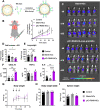
Antitumoral efficacy of poly(I:C)+R848-HA-man-prot-NCs after intravenous injection in the orthotopic lung cancer murine model CMT167. (A) Schematic representation of the coating of the pIC+R848-protamine-NCs with HA-mannose (here abbreviated as pIC+R848-NCs), and description of their components. (B) Evolution of luminescence/tumor signal of CMT167-Luc tumor-bearing mice treated with the NCs (3 intravenous injections corresponding to 25 µg of each drug at times indicated by arrows). (C) Representative pictures of the luminescence signal in the whole animal at 8, 18 and 22 days. (D) Comparison of the tumor signals in each treatment group at sacrifice. (E) Comparison of the lungs weights at sacrifice. (F) Quantification of circulation levels of CXCL10, TNF-α and IL-6 in the peripheral blood collected at day 18. (G) Body weight of CMT167-Luc tumor-bearing mice treated with the NCs (3 intravenous injections corresponding to 25 µg of each drug at times as indicated in Figure 5) along the whole experiment (average of 6 mice), and (H) last measurement at day 22. (I) Spleen weight at sacrifice (day 22) (each point represents 1 mouse). Values represent mean ± s.e.m. Statistical comparison was performed using a one-way ANOVA followed by a Tukey’s multiple comparison test. Statistically significant differences are represented as *(p<0.05), **(p<0.01) and ****(p<0.0001). pIC, poly(I:C); R848, resiquimod. pIC+R848-NCs, pIC+R848-protamine-NCs with HA-mannose. Blank-NCs, equivalent nanoformulations, coated with HA-mannose, but not loaded with the drugs pIC+R848.
Figure 6
Figure 6
Tumor microenvironment analysis, with a focus on TAMs, after intravenous injection of poly(I:C)+R848-HA-man-prot-NCs in the orthotopic lung cancer murine model CMT167. (A) Flow cytometry analysis of single-cell suspensions of tumor-bearing lungs: total leukocytes (CD45+) among living cells, and quantification of interstitial macrophages (CD64+; Cd11bhigh) among total macrophages, and CD206 expression; quantification of alveolar macrophages (CD64+; Cd11blow) among total macrophages, and CD206 expression. (B, C) Multiplexed immunofluorescence analysis of CMT167-Luc-derived tumors at sacrifice. Quantification of CD8+ cells, CD4+ cells, FOXP3+/CD4+ Treg cells and ratio of M1 (F4/80+, CD86+):M2 (F4/80+, Arg1+) macrophages in treated tumors. (C) Representative images of controls, tumors treated with blank-NCs or with pIC+R848-NCs. DAPI was used for staining of nucleus, F4/80 for macrophages, CD86 as M1 marker and Arg1 as M2 marker. Values represent mean ± s.e.m. Statistical comparison was performed using a one-way ANOVA followed by a Tukey’s multiple comparison test. Statistically significant differences are represented as *(p<0.05) and **(p<0.01). pIC, poly(I:C); R848, resiquimod. pIC+R848-NCs, pIC+R848-protamine-NCs with HA-mannose. Blank-NCs, equivalent nanoformulations, coated with HA-mannose, but not loaded with the drugs pIC+R848.

References

    1. Mantovani A, Allavena P, Sica A, Balkwill F. Cancer-related inflammation. Nature (2008) 454:436–44. doi: 10.1038/nature07205 - DOI - PubMed
    1. Hanahan D, Weinberg RA. Hallmarks of cancer: the next generation. Cell (2011) 144:646–74. doi: 10.1016/j.cell.2011.02.013 - DOI - PubMed
    1. Mantovani A, Marchesi F, Malesci A, Laghi L, Allavena P. Tumour-associated macrophages as treatment targets in oncology. Nat Rev Clin Oncol (2017) 14:399–416. doi: 10.1038/nrclinonc.2016.217 - DOI - PMC - PubMed
    1. Cassetta L, Pollard JW. A timeline of tumour-associated macrophage biology. Nat Rev Cancer (2023) 23:238–57. doi: 10.1038/s41568-022-00547-1 - DOI - PubMed
    1. Allavena P, Anfray C, Ummarino A, Andón FT. Therapeutic manipulation of tumor-associated macrophages: facts and hopes from a clinical and translational perspective. Clin Cancer Res (2021) 27:3291–7. doi: 10.1158/1078-0432.CCR-20-1679 - DOI - PubMed

Publication types