

Autophagy in spinal muscular atrophy: from pathogenic mechanisms to therapeutic approaches
- PMID: 38259504
- PMCID: PMC10801191
- DOI: 10.3389/fncel.2023.1307636
Autophagy in spinal muscular atrophy: from pathogenic mechanisms to therapeutic approaches
Abstract
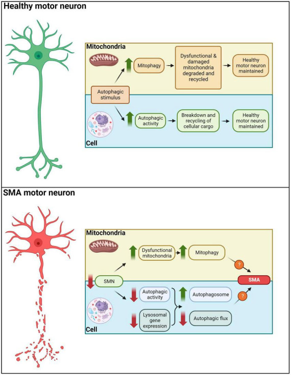
Spinal muscular atrophy (SMA) is a devastating neuromuscular disorder caused by the depletion of the ubiquitously expressed survival motor neuron (SMN) protein. While the genetic cause of SMA has been well documented, the exact mechanism(s) by which SMN depletion results in disease progression remain elusive. A wide body of evidence has highlighted the involvement and dysregulation of autophagy in SMA. Autophagy is a highly conserved lysosomal degradation process which is necessary for cellular homeostasis; defects in the autophagic machinery have been linked with a wide range of neurodegenerative disorders, including amyotrophic lateral sclerosis, Alzheimer's disease and Parkinson's disease. The pathway is particularly known to prevent neurodegeneration and has been suggested to act as a neuroprotective factor, thus presenting an attractive target for novel therapies for SMA patients. In this review, (a) we provide for the first time a comprehensive summary of the perturbations in the autophagic networks that characterize SMA development, (b) highlight the autophagic regulators which may play a key role in SMA pathogenesis and (c) propose decreased autophagic flux as the causative agent underlying the autophagic dysregulation observed in these patients.
Keywords: autophagic flux; autophagy; macroautophagy; mitophagy; spinal muscular atrophy.
Copyright © 2024 Rashid and Dimitriadi.
Conflict of interest statement
The authors declare that the research was conducted in the absence of any commercial or financial relationships that could be construed as a potential conflict of interest.
Figures
Similar articles
-
Survival motor neuron protein reduction deregulates autophagy in spinal cord motoneurons in vitro.Cell Death Dis. 2013 Jun 20;4(6):e686. doi: 10.1038/cddis.2013.209. Cell Death Dis. 2013. PMID: 23788043 Free PMC article.
-
Autophagy dysregulation in cell culture and animals models of spinal muscular atrophy.Mol Cell Neurosci. 2014 Jul;61:133-40. doi: 10.1016/j.mcn.2014.06.006. Epub 2014 Jun 28. Mol Cell Neurosci. 2014. PMID: 24983518 Free PMC article.
-
Spinal Muscular Atrophy autophagy profile is tissue-dependent: differential regulation between muscle and motoneurons.Acta Neuropathol Commun. 2021 Jul 3;9(1):122. doi: 10.1186/s40478-021-01223-5. Acta Neuropathol Commun. 2021. PMID: 34217376 Free PMC article.
-
Therapy development for spinal muscular atrophy: perspectives for muscular dystrophies and neurodegenerative disorders.Neurol Res Pract. 2022 Jan 4;4(1):2. doi: 10.1186/s42466-021-00162-9. Neurol Res Pract. 2022. PMID: 34983696 Free PMC article. Review.
-
Modelling motor neuron disease in fruit flies: Lessons from spinal muscular atrophy.J Neurosci Methods. 2018 Dec 1;310:3-11. doi: 10.1016/j.jneumeth.2018.04.003. Epub 2018 Apr 9. J Neurosci Methods. 2018. PMID: 29649521 Review.
Cited by
-
CK and LRRK2 Involvement in Neurodegenerative Diseases.Int J Mol Sci. 2024 Oct 30;25(21):11661. doi: 10.3390/ijms252111661. Int J Mol Sci. 2024. PMID: 39519213 Free PMC article. Review.
-
The role of NLRP3 inflammasome in multiple sclerosis: pathogenesis and pharmacological application.Front Immunol. 2025 Apr 2;16:1572140. doi: 10.3389/fimmu.2025.1572140. eCollection 2025. Front Immunol. 2025. PMID: 40242770 Free PMC article. Review.
-
Real-World Safety Data of the Orphan Drug Onasemnogene Abeparvovec (Zolgensma®) for the SMA Rare Disease: A Pharmacovigilance Study Based on the EMA Adverse Event Reporting System.Pharmaceuticals (Basel). 2024 Mar 19;17(3):394. doi: 10.3390/ph17030394. Pharmaceuticals (Basel). 2024. PMID: 38543180 Free PMC article.
-
[Research progress on phenotypic modifier genes in spinal muscular atrophy].Zhongguo Dang Dai Er Ke Za Zhi. 2025 Feb 15;27(2):229-235. doi: 10.7499/j.issn.1008-8830.2410064. Zhongguo Dang Dai Er Ke Za Zhi. 2025. PMID: 39962788 Free PMC article. Review. Chinese.
-
Significance of Programmed Cell Death Pathways in Neurodegenerative Diseases.Int J Mol Sci. 2024 Sep 15;25(18):9947. doi: 10.3390/ijms25189947. Int J Mol Sci. 2024. PMID: 39337436 Free PMC article. Review.
References
-
- Axe E., Walker S., Manifava M., Chandra P., Roderick H., Habermann A., et al. (2008). Autophagosome formation from membrane compartments enriched in phosphatidylinositol 3-phosphate and dynamically connected to the endoplasmic reticulum. J. Cell Biol. 182 685–701. 10.1083/jcb.200803137 - DOI - PMC - PubMed
-
- Barrett D., Bilic S., Chyung Y., Cote S., Iarrobino R., Kacena K., et al. (2021). A randomized phase 1 safety, pharmacokinetic and pharmacodynamic study of the novel myostatin inhibitor apitegromab (SRK-015): a potential treatment for spinal muscular atrophy. Adv. Ther. 38 3203–3222. 10.1007/s12325-021-01757-z - DOI - PMC - PubMed
Publication types
LinkOut - more resources
Full Text Sources