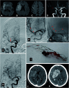
Symptomatic Intracranial Hemorrhage after Mechanical Thrombectomy - the Difference between Iso-Osmolar and Low-Osmolar Contrast Media
- PMID: 38260038
- PMCID: PMC10800169
- DOI: 10.5797/jnet.oa.2023-0074
Symptomatic Intracranial Hemorrhage after Mechanical Thrombectomy - the Difference between Iso-Osmolar and Low-Osmolar Contrast Media
Abstract
Objective: Symptomatic intracranial hemorrhage (SICH) after mechanical thrombectomy (MT) is generally considered a critical complication. Hemorrhagic transformation after ischemic stroke has also been associated with contrast media administration. The objective of our study was to evaluate correlations between contrast media type and incidence of SICH after MT.
Methods: Ninety-three consecutive patients (41 men; mean age, 80.2 years; range, 44-98 years) underwent MT reperfusion (expanded thrombolysis in cerebral infarction score, 2a-3) for acute large-vessel occlusion ischemic stroke within 8 h after symptom onset between April 2020 and July 2023 were retrospectively reviewed. Correlations between contrast media type (iso-osmolar or low-osmolar medium) and incidence of SICH were assessed.
Results: Contrast media were iso-osmolar in 60 cases or low-osmolar in 33 cases. The overall incidence of SICH was 5.5%. The frequency of SICH was significantly lower in the iso-osmolar group (1.7%) than in the low-osmolar group (12.1%; P = 0.033).
Conclusion: Iso-osmolar contrast media was associated with a lower incidence of SICH compared with low-osmolar contrast media in patients after MT.
Keywords: iso-osmolar contrast media; mechanical thrombectomy; symptomatic intracranial hemorrhage.
©2024 The Japanese Society for Neuroendovascular Therapy.
Figures


Similar articles
-
Hemorrhagic Transformation Rates following Contrast Media Administration in Patients Hospitalized with Ischemic Stroke.AJNR Am J Neuroradiol. 2022 Mar;43(3):381-387. doi: 10.3174/ajnr.A7412. Epub 2022 Feb 10. AJNR Am J Neuroradiol. 2022. PMID: 35144934 Free PMC article.
-
External validation of TICI-ASPECTS-glucose score as a predictive model for symptomatic intracranial hemorrhage following mechanical thrombectomy.J Stroke Cerebrovasc Dis. 2022 Nov;31(11):106796. doi: 10.1016/j.jstrokecerebrovasdis.2022.106796. Epub 2022 Sep 29. J Stroke Cerebrovasc Dis. 2022. PMID: 36183517
-
Contrast Neurotoxicity and its Association with Symptomatic Intracranial Hemorrhage After Mechanical Thrombectomy.Clin Neuroradiol. 2022 Dec;32(4):961-969. doi: 10.1007/s00062-022-01152-3. Epub 2022 Mar 16. Clin Neuroradiol. 2022. PMID: 35294573
-
Comparison of Risk Factors, Safety, and Efficacy Outcomes of Mechanical Thrombectomy in Posterior vs. Anterior Circulation Large Vessel Occlusion.Front Neurol. 2021 Jun 22;12:687134. doi: 10.3389/fneur.2021.687134. eCollection 2021. Front Neurol. 2021. PMID: 34239498 Free PMC article.
-
Symptomatic intracranial hemorrhage following intravenous thrombolysis for acute ischemic stroke: a critical review of case definitions.Cerebrovasc Dis. 2012;34(2):106-14. doi: 10.1159/000339675. Epub 2012 Aug 1. Cerebrovasc Dis. 2012. PMID: 22868870 Review.
Cited by
-
A clinical and computed tomography-based nomogram to predict the outcome in patients with anterior circulation large vessel occlusion after endovascular mechanical thrombectomy.Jpn J Radiol. 2024 Sep;42(9):973-982. doi: 10.1007/s11604-024-01583-7. Epub 2024 May 3. Jpn J Radiol. 2024. PMID: 38700623
References
-
- Goyal M, Menon BK, van Zwam WH, et al. Endovascular thrombectomy after large-vessel ischaemic stroke: a meta-analysis of individual patient data from five randomised trials. Lancet 2016; 387: 1723–1731. - PubMed
-
- Hao Y, Yang D, Wang H, et al. Predictors for symptomatic intracranial hemorrhage after endovascular treatment of acute ischemic stroke. Stroke 2017; 48: 1203–1209. - PubMed
-
- Zhang X, Xie Y, Wang H, et al. Symptomatic intracranial hemorrhage after mechanical thrombectomy in Chinese ischemic stroke patients. Stroke 2020; 51: 2690–2696. - PubMed
LinkOut - more resources
Full Text Sources
Miscellaneous

