[Preprint]. 2024 Jan 9:2024.01.08.24301002.
doi: 10.1101/2024.01.08.24301002.

Immunological imprinting shapes the specificity of human antibody responses against SARS-CoV-2 variants

Affiliations

Immunological imprinting shapes the specificity of human antibody responses against SARS-CoV-2 variants

Timothy S Johnston et al. medRxiv. .

Update in

Abstract

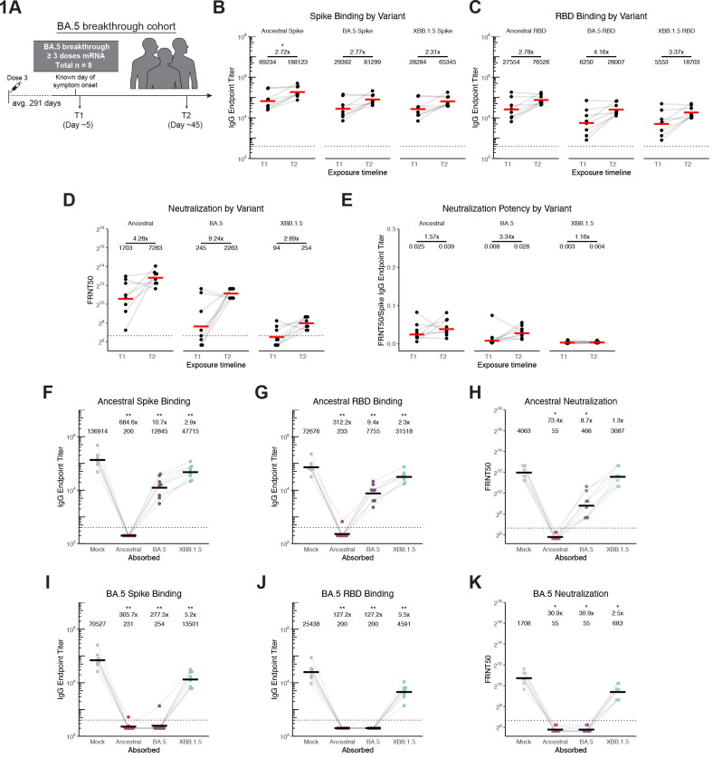
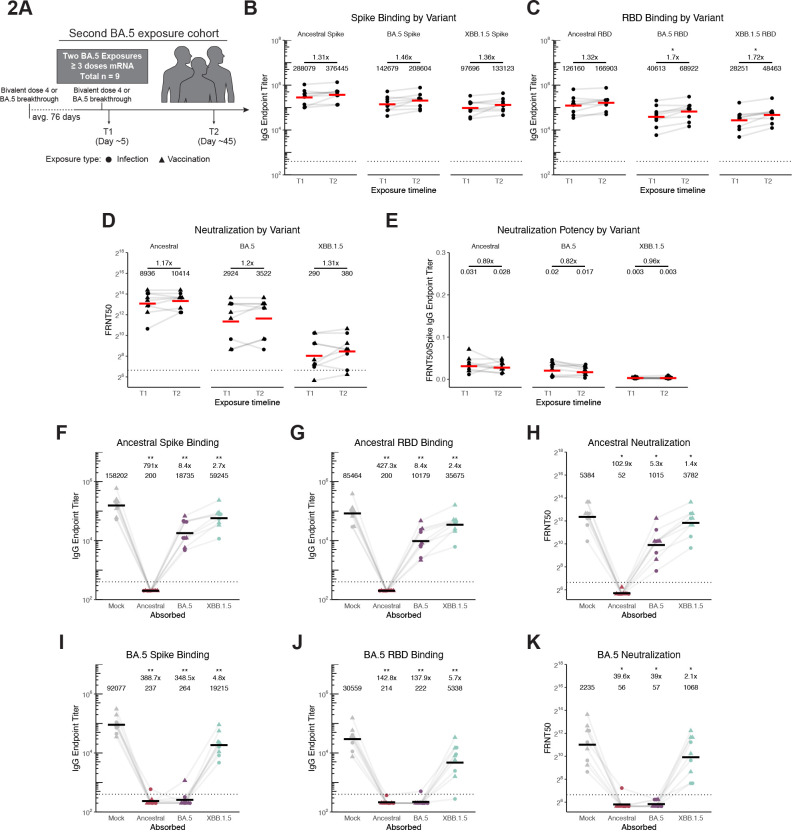
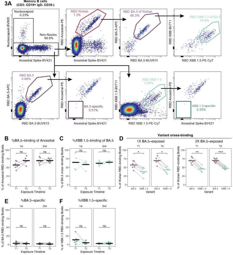
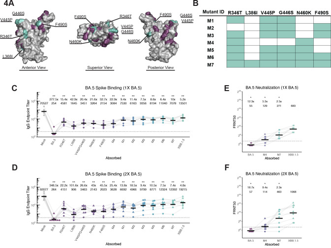
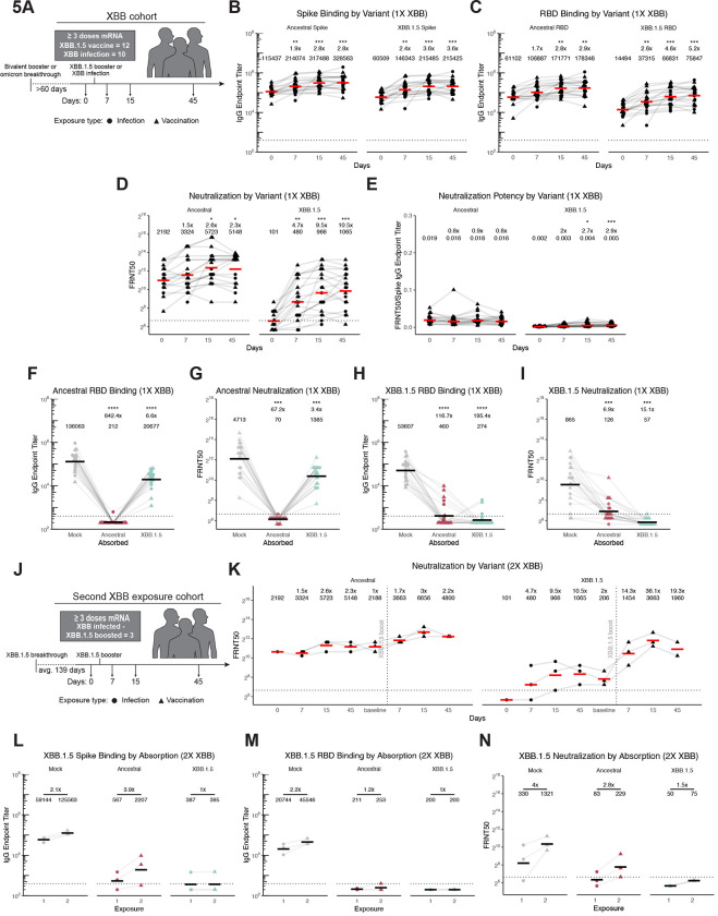
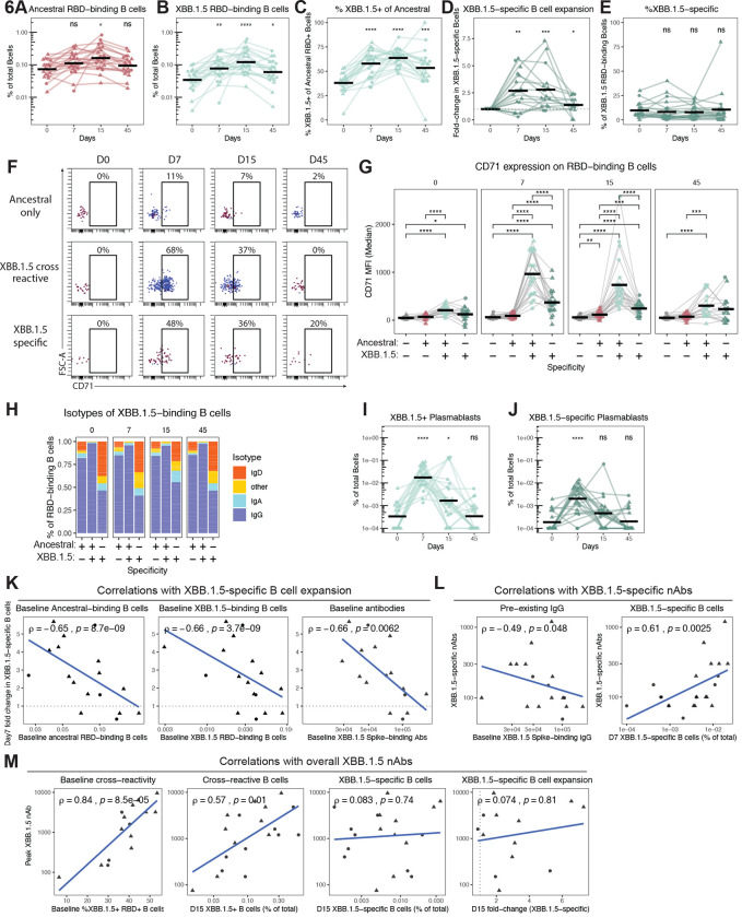
The spike glycoprotein of severe acute respiratory syndrome coronavirus-2 (SARS-CoV-2) continues to accumulate substitutions, leading to breakthrough infections of vaccinated individuals and prompting the development of updated booster vaccines. Here, we determined the specificity and functionality of antibody and B cell responses following exposure to BA.5 and XBB variants in individuals who received ancestral SARS-CoV-2 mRNA vaccines. BA.5 exposures elicited antibody responses that primarily targeted epitopes conserved between the BA.5 and ancestral spike, with poor reactivity to the XBB.1.5 variant. XBB exposures also elicited antibody responses that targeted epitopes conserved between the XBB.1.5 and ancestral spike. However, unlike BA.5, a single XBB exposure elicited low levels of XBB.1.5-specific antibodies and B cells in some individuals. Pre-existing cross-reactive B cells and antibodies were correlated with stronger overall responses to XBB but weaker XBB-specific responses, suggesting that baseline immunity influences the activation of variant-specific SARS-CoV-2 responses.

Keywords: B cells; Omicron BA.5; SARS-CoV-2; XBB.1.5; antibodies; immune evasion; immune imprinting; mRNA vaccines; original antigenic sin; viral evolution.


Conflict of interest statement

Declaration of Interests E.J.W. is a member of the Parker Institute for Cancer Immunotherapy. E.J.W. is an advisor for Arsenal Biosciences, Coherus, Danger Bio, IpiNovyx, Janssen, New Limit, Marengo, Pluto Immunotherapeutics Related Sciences, Santa Ana Bio, and Synthekine. E.J.W. is a founder of and holds stock in Coherus, Danger Bio, and Arsenal Biosciences.

Figures

Figure 1.
Figure 1.
BA.5 breakthrough infection elicits cross-reactive antibodies evaded by XBB.1.5. A) Schematic of participants in this study who were vaccinated 3x with the SARS-CoV-2 ancestral mRNA-LNP vaccine who then had a BA.5 breakthrough infection. B)-C) Antigen-specific IgG ELISAs were performed using sera from timepoints indicated in panel A) against ancestral, BA.5, and XBB.1.5 full-length spike (B) and RBD (C) proteins. Endpoint titers are reported as reciprocal serum dilutions. D) SARS-CoV-2 pseudotype neutralization assays were performed using sera obtained at timepoints indicated in panel A) against ancestral SARS-CoV-2, BA.5, and XBB.1.5 pseudoviruses. Values reported are focus reduction neutralization test (FRNT) 50, or reciprocal serum dilution at which <50% viral input foci are observed. E) Neutralization potency was calculated by dividing FRNT50 values by spike IgG titer. F-G) Antigen-specific IgG ELISAs were performed using T2 absorbed sera and ELISA plates coated with the ancestral spike (F) or ancestral RBD (G). H) SARS-CoV-2 pseudotype neutralization assays were performed using T2 absorbed serum and ancestral SARS-CoV-2 pseudovirus. I-J) Antigen-specific IgG ELISAs were performed using T2 absorbed and ELISA plates coated with BA.5 spike (I) and BA.5 RBD (J). K) Neutralization assays were performed using T2 absorbed serum and BA.5 pseudotyped virus. For all, individual points are average of n = 2 technical replicates. Red/black bars indicate geometric mean. Wilcoxon signed-rank test with benjamini-hochberg correction for multiple testing. All comparisons to timepoint 1 or mock absorption. * p<0.05, ** p<0.01.
Figure 2.
Figure 2.
Secondary BA.5 exposure elicits cross-reactive antibodies that bind weakly to XBB.1.5. A) Schematic of participants in this study who were vaccinated 3x with the SARS-CoV-2 ancestral mRNA-LNP vaccine who then had two BA.5 exposures. B)-C) Antigen-specific IgG ELISAs were performed using sera from timepoints indicated in panel A) against ancestral, BA.5, and XBB.1.5 full-length spike (B) and RBD (C). Endpoint titers are reported as reciprocal dilutions. D) SARS-CoV-2 pseudotype neutralization assays were performed using sera obtained at timepoints indicated in panel A) against ancestral SARS-CoV-2, BA.5, and XBB.1.5 pseudoviruses. Values reported are focus reduction neutralization test (FRNT) 50, or reciprocal serum dilution at which <50% viral input foci are observed. E) Neutralization potency was calculated by dividing FRNT50 values by spike IgG titer. F)-G) Antigen-specific IgG ELISAs were performed using T2 absorbed sera and ELISA plates coated with the ancestral spike (F) or ancestral RBD (G). H) SARS-CoV-2 pseudotype neutralization assays were performed using T2 absorbed serum and ancestral SARS-CoV-2 pseudovirus. I-J) Antigen-specific IgG ELISAs were performed using T2 absorbed sera and ELISA plates coated with BA.5 spike (I) and BA.5 RBD (J). K) Neutralization assays were performed using T2 absorbed serum and BA.5 pseudotyped virus. For all, Individual points are average of n = 2 technical replicates. Red/black bars indicate geometric mean. Wilcoxon signed-rank test with benjamini-hochberg correction for multiple testing. All comparisons to timepoint 1 or mock absorption. * p<0.05, ** p<0.01.
Figure 3.
Figure 3.
Cross-reactive B cells are recruited in response to primary and secondary BA.5 exposures in vaccinated individuals. A) Representative flow-cytometry plot showing gating strategy for probe positive memory B cells. B) The percent of ancestral RBD-binding B cells that cross-bind to BA.5 RBD before and after first (left) and second (right) exposure to BA.5. C) The percent of ancestral RBD and BA.5 RBD cross-binding B cells that also cross-bind to XBB.1.5 RBD before and after first (left) and second (right) exposure to BA.5. D) The percent of ancestral RBD-binding B cells that cross-bind BA.5 and XBB.1.5 RBD before and after first (left) and second (right) exposure to BA.5. E-F) The frequency of BA.5 RBD-binding (E) and XBB.1.5 RBD-binding (F) B cells that do not cross-react with ancestral RBD before and after first (left) and second (right) exposure to BA.5.
Figure 4.
Figure 4.
Antibodies elicited by successive ancestral SARS-CoV-2 and BA.5 exposures target conserved residues that are mutated in the XBB.1.5 RBD. A) Ancestral RBD structure with residues that are different in BA.5 are highlighted in purple, and residues conserved between ancestral and BA.5 but different in XBB.1.5 are highlighted in teal (PDB 6M0J). B) List of recombinant mutations created for this study using the BA.5 spike backbone. C) IgG ELISA using T2 breakthrough sera after absorption with single mutants and those listed in panel B. D) IgG ELISA usingT2 sera from individuals exposed twice with BA.5 after absorption with single mutants and those listed in panel B. E,F) Neutralization assays were performed using T2 absorbed serum from single (E) and two (F) BA.5 exposures using BA.5 pseudotype virus. For all, individual points are average of n = 2 technical replicates. Black bars indicate geometric mean. Wilcoxon signed-rank test with benjamini-hochberg correction for multiple testing. All comparisons to mock absorption. * p<0.05, ** p<0.01.
Figure 5.
Figure 5.
Primary and secondary XBB exposures elicit mostly antibodies that cross-react to the ancestral SARS-CoV-2 spike. A) Schematic of participants in this study who were exposed to XBB. B)-C) Antigen-specific IgG ELISAs were performed using sera from timepoints indicated in panel A) against ancestral and XBB.1.5 full-length spike (B) and RBD (C). Endpoint titers are reported as reciprocal dilutions. D) Neutralization assays were performed using serum obtained at timepoints indicated in panel A) against ancestral and XBB.1.5 SARS-CoV-2 pseudoviruses. Values reported are focus reduction neutralization test (FRNT) 50, or reciprocal serum dilution at which <50% viral input foci are observed. E) Neutralization potency was calculated by dividing FRNT50 values by spike IgG titer. F),H) Antigen-specific IgG ELISAs were performed using sera (collected 45 days after XBB1.5 exposure) after absorption with ancestral or XBB.1.5 spike proteins. G),I) SARS-CoV-2 pseudotype neutralization assays were performed using sera (collected 45 days after XBB.1.5 exposure) after absorption with ancestral or XBB.1.5 spike proteins. J) Schematic of participants in this study who were exposed twice to XBB. K) SARS-CoV-2 pseudotype neutralization assays were performed using sera obtained at timepoints indicated in panel J) using ancestral and XBB.1.5 pseudoviruses. L),M) Antigen-specific IgG ELISAs and N) SARS-CoV-2 neutralization assays were performed using sera (collected 45 days after each XBB.1.5 exposure) after absorption with ancestral or XBB1.5 spike proteins. For all, Individual points are average of n = 2 technical replicates. Red/black bars indicate geometric mean. Wilcoxon rank-sum test with benjamini-hochberg correction for multiple testing. All comparisons to timepoint 1 or mock absorption. * p<0.05, ** p<0.01, *** p<0.001, **** p<0.0001.
Figure 6.
Figure 6.
Cross-reactive B cells dominate immune responses elicited by XBB, but variant-specific responses are observed in individuals with low pre-existing immunity. A-B) The percent of total B cells that bind A) ancestral RBD and B) XBB.1.5 RBD before and after exposure to XBB.1.5. C) The percent of ancestral RBD-binding B cells that cross-bind to XBB.1.5 RBD before and after exposure to XBB.1.5. D) The fold expansion of B cells that bind XBB.1.5 RBD but not ancestral RBD (XBB.1.5-specific) following XBB.1.5 exposure. E) The percent of total XBB.1.5 RBD-binding B cells that do not bind ancestral RBD. F) Representative flow cytometry plots depicting longitudinal changes in CD71 expression on different RBD-binding B cell populations from a single individual. G) Summary data of median CD71 expression on the indicated RBD-binding B cell populations at 0, 7, 15, and 45 days post-XBB exposure. H) Summary data of the isotype distribution of RBD-binding B cells at 0, 7, 15, and 45 days post-XBB exposure. I-J) The percent of total B cells that are CD38+CD27+ plasmablasts and I) bind XBB.1.5 RBD or J) bind XBB.1.5 RBD and do not bind ancestral RBD (XBB.1.5-specific). K-M) Correlations within B cell and antibody responses. K) Correlation of day 7 fold change in XBB.1.5-specific B cells as in panel D with day 0 percent of total B cells that bind ancestral RBD (left); day 0 percent of total B cells that bind XBB.1.5 RBD (center); day 0 XBB.1.5 spike-binding antibodies by ELISA (right). L) Correlations of ancestral spike absorbed XBB.1.5 neutralizing antibody titers at day 45 (as in Fig. 5N) with day 0 XBB.1.5 spike-binding antibodies quantified by ELISA (left), and the percent of total B cells that bind XBB.1.5 RBD but not ancestral RBD at day 7 (right). M) Correlation of overall day 45 XBB.1.5 neutralizing antibody titers (unabsorbed) with the day 0 percent of ancestral RBD+ B cells that cross-bind XBB.1.5 RBD; the day 15 percentage of total B cells that bind XBB.1.5 RBD, the day 15 percentage of total B cells that bind XBB.1.5 RBD but do not bind ancestral RBD (XB.1.5-specific), and the day 15 fold change in XBB-specific B cells as in D. Points represent individual subjects and thin lines indicate individual subjects sampled longitudinally. Horizontal bars represent means. Statistics were calculated using two-sided Wilcoxon test with Benjamini-Hochberg correction for multiple comparisons. Statistics without brackets are in comparison to day 0. Correlation statistics were calculated using Spearman rank correlation and are shown with Pearson trend lines for visualization.

Similar articles

References

    1. Corbett K.S., Edwards D.K., Leist S.R., Abiona O.M., Boyoglu-Barnum S., Gillespie R. A., Himansu S., Schäfer A., Ziwawo C.T., DiPiazza A.T., Dinnon K.H., Elbashir S. M., Shaw C.A., Woods A., Fritch E.J., Martinez D.R., Bock K.W., Minai M., Nagata B.M., Hutchinson G.B., Wu K., Henry C., Bahl K., Garcia-Dominguez D., Ma L., Renzi I., Kong W.-P., Schmidt S.D., Wang L., Zhang Y., Phung E., Chang L.A., Loomis R.J., Altaras N.E., Narayanan E., Metkar M., Presnyak V., Liu C., Louder M. K., Shi W., Leung K., Yang E.S., West A., Gully K.L., Stevens L.J., Wang N., Wrapp D., Doria-Rose N.A., Stewart-Jones G., Bennett H., Alvarado G.S., Nason M.C., Ruckwardt T.J., McLellan J.S., Denison M.R., Chappell J.D., Moore I.N., Morabito K.M., Mascola J.R., Baric R.S., Carfi A., Graham B.S., 2020. SARS-CoV-2 mRNA vaccine design enabled by prototype pathogen preparedness. Nature 586, 567–571. 10.1038/s41586-020-2622-0 - DOI - PMC - PubMed
    1. Hsieh C.-L., Goldsmith J.A., Schaub J.M., DiVenere A.M., Kuo H.-C., Javanmardi K., Le K.C., Wrapp D., Lee A.G., Liu Y., Chou C.-W., Byrne P.O., Hjorth C.K., Johnson N. V., Ludes-Meyers J., Nguyen A.W., Park J., Wang N., Amengor D., Lavinder J.J., Ippolito G.C., Maynard J.A., Finkelstein I.J., McLellan J.S., 2020. Structure-based design of prefusion-stabilized SARS-CoV-2 spikes. Science eabd0826. 10.1126/science.abd0826 - DOI - PMC - PubMed
    1. Jackson L.A., Anderson E.J., Rouphael N.G., Roberts P.C., Makhene M., Coler R.N., McCullough M.P., Chappell J.D., Denison M.R., Stevens L.J., Pruijssers A.J., McDermott A., Flach B., Doria-Rose N.A., Corbett K.S., Morabito K.M., O’Dell S., Schmidt S.D., Swanson P.A., Padilla M., Mascola J.R., Neuzil K.M., Bennett H., Sun W., Peters E., Makowski M., Albert J., Cross K., Buchanan W., Pikaart-Tautges R., Ledgerwood J.E., Graham B.S., Beigel J.H., mRNA-1273 Study Group, 2020. An mRNA Vaccine against SARS-CoV-2 - Preliminary Report. N Engl J Med 383, 1920–1931. 10.1056/NEJMoa2022483 - DOI - PMC - PubMed
    1. Mannar D., Saville J.W., Zhu X., Srivastava S.S., Berezuk A.M., Tuttle K.S., Marquez A.C., Sekirov I., Subramaniam S., 2022. SARS-CoV-2 Omicron variant: Antibody evasion and cryo-EM structure of spike protein–ACE2 complex. Science 375, 760–764. 10.1126/science.abn7760 - DOI - PMC - PubMed
    1. Accorsi E.K., Britton A., Fleming-Dutra K.E., Smith Z.R., Shang N., Derado G., Miller J., Schrag S.J., Verani J.R., 2022. Association Between 3 Doses of mRNA COVID-19 Vaccine and Symptomatic Infection Caused by the SARS-CoV-2 Omicron and Delta Variants. JAMA 327, 639–651. 10.1001/jama.2022.0470 - DOI - PMC - PubMed

Publication types