[Preprint]. 2024 Jul 12:2024.01.02.573939.
doi: 10.1101/2024.01.02.573939.

Development of robust antiviral assays using relevant apical-out human airway organoids

Affiliations

Development of robust antiviral assays using relevant apical-out human airway organoids

Ji-Hoon Lee et al. bioRxiv. .

Abstract

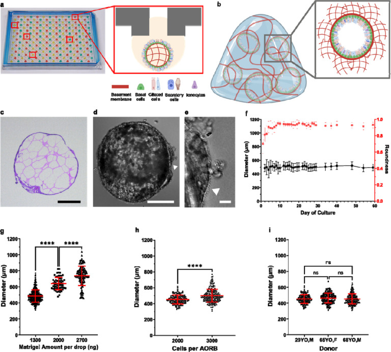
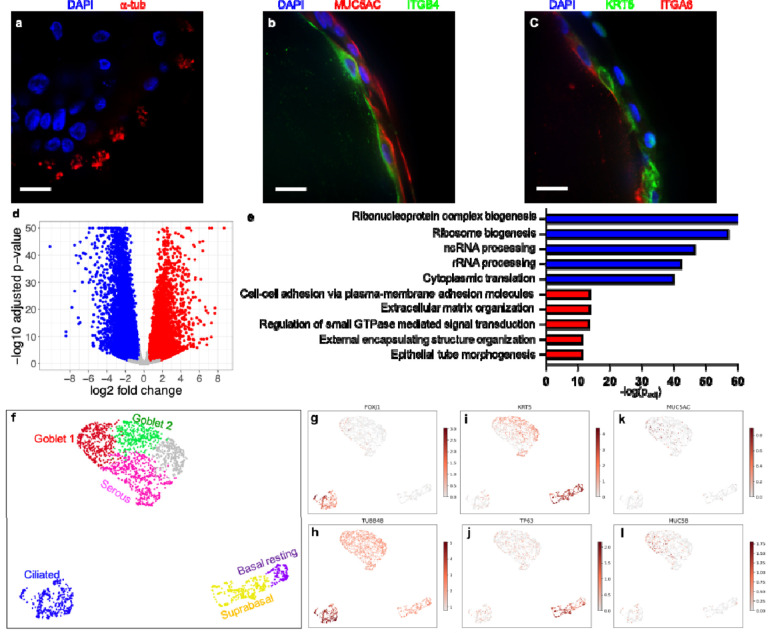
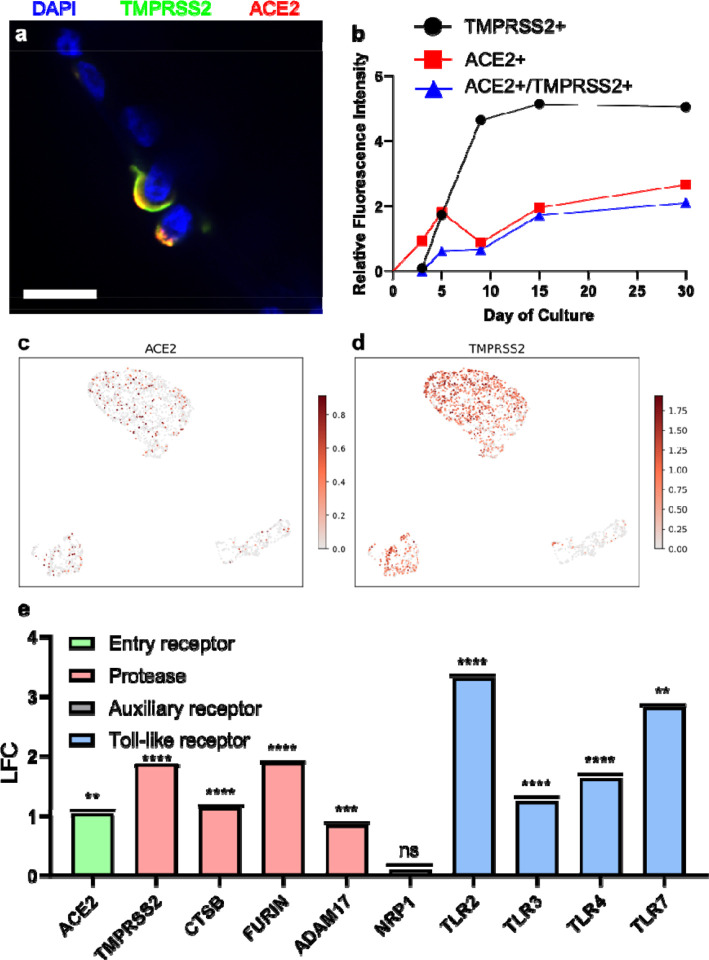
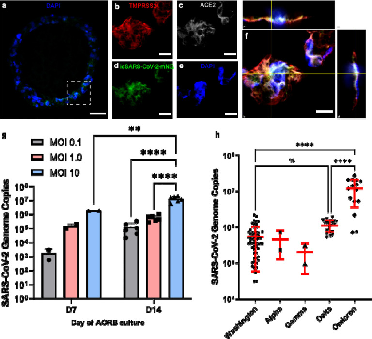
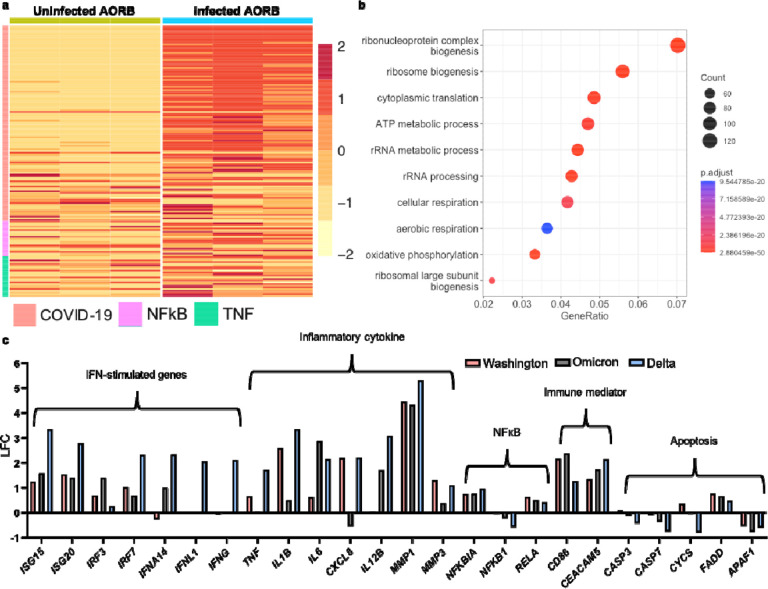
While breakthroughs with organoids have emerged as next-generation in vitro tools, standardization for drug discovery remains a challenge. This work introduces human airway organoids with reversed biopolarity (AORBs), cultured and analyzed in a high-throughput, single-organoid-per-well format, enabling milestones towards standardization. AORBs exhibit a spatio-temporally stable apical-out morphology, facilitating high-yield direct intact-organoid virus infection. Single-cell RNA sequencing and immunohistochemistry confirm the physiologically relevant recapitulation of differentiated human airway epithelia. The cellular tropism of five severe acute respiratory syndrome coronavirus 2 (SARS-CoV-2) strains along with host response differences between Delta, Washington, and Omicron variants, as observed in transcriptomic profiles, also suggest clinical relevance. Dose-response analysis of three well-studied SARS-CoV-2 antiviral compounds (remdesivir, bemnifosbuvir, and nirmatrelvir) demonstrates that AORBs efficiently predict human efficacy, comparable to gold-standard air-liquid interface cultures, but with higher throughput (~10-fold) and fewer cells (~100-fold). This combination of throughput and relevance allows AORBs to robustly detect false negative results in efficacy, preventing irretrievable loss of promising lead compounds. While this work leverages the SARS-CoV-2 study as a proof-of-concept application, the standardization capacity of AORB holds broader implications in line with regulatory efforts to push alternatives to animal studies.

Keywords: AT-511; Organoid; SARS-CoV-2; antiviral testing; apical-out; high-throughput; nirmatrelvir; remdesivir; standardization.


Conflict of interest statement

Competing Interests Lee, Parigoris, and Takayama are inventors on US20230194506A1, filed on May 21, 2021, related to this work.

Figures

Fig. 1|
Fig. 1|. Human airway epithelial organoid with reversed biopolarity (AORBs).
a, A 192 hanging drop array culture of single AORB-per-drop provides throughput and consistency. Eversion-free stably-inverted AORBs have terminally differentiated and physiologically relevant cell types. b, Conventional Matrigel-dome method of organoid culture has an exterior basement membrane with the apical side facing the interior. c, H&E staining shows an epithelial layer shell with a core filled with Matrigel. d, Phase-contrast image of an entire AORB, and e, a zoomed-in image showing cilia (denoted by triangles) and AORB-produced mucus. f, The diameters of AORBs are stable over 80 days, whereas the roundness increases up to day 10 of culture then stabilizes, n = 16 AORBs per timepoint. g, For a given number of starting cells, Matrigel amount can alter AORB diameters, n = 434, 66, 116 individual AORBs for 1300, 2000, and 2700 ng, respectively. h, Seeding density also provides a statistically significant means of regulating AORB diameters, n = 147 and 287 AORBs for 2000 and 3000 seeding cells, respectively. i, For a given Matrigel amount and cell density, AORB diameters are consistent across biological donors, n = 147, 157, and 290 for the three donors, respectively. ns, p>0.05, ****p<0.0001. Error bars indicate the mean ± 1 standard deviation. Scale bars, 250 μm in c and d, and 20 μm in e.
Fig. 2|
Fig. 2|. Confocal Immunofluorescence imaging confirms heterogeneous cell population and apical-out polarization in AORBs.
a, Acetylated alpha tubulin orienting outwards indicating that the apical side of the ciliated cells face the organoid exterior. b, AORBs also have positive MUC5AC (red) indicative of goblet cells. ITGB4 (green) binds the basement membrane and indicates the basolateral side is facing the organoid interior. c, KRT5 (green) and ITGA6 (red) stains indicate the presence of basal cells. d, Volcano plot from bulk RNA sequencing comparing transcriptomic profile of the differentiated AORBs with respect to the initial cells before seeding (2D cultures) shows 7521 upregulated and 7100 downregulated genes. e, Top 5 statistically significant upregulated (red) and downregulated (blue) biological processes from gene ontology analysis of the enriched gene sets from DGE analysis. f, UMAP of scRNAseq results (n = 2651 cells) of AORBs showing three main clusters, ciliated, basal, and secretory. Further clustering shows sub-cell types, reprehensive of human bronchi. g-l, Canonical markers of ciliated cells (g and h), basal cells (i and j), and goblet cells (k and l). Color bars represent log-transformed normalized expression. Scale bars, 20 μm, in a-c.
Fig. 3|
Fig. 3|. IL-13-induced apical-out mucus hypersecretion experiment.
a, Apical-out morphology in AORBs allows efficient assay to study mucus secretion. b and c, Bright-field and immunofluorescent images of an AORB treated with 1 ng/mL IL-13, respectively. Nuclei and mucus are stained with Hoechst 33342 (blue) and wheat germ agglutinin (green). d, Representative bright-field images of untreated AORBs. e, Representative bright-field images of IL-13-treated. f, DGE analysis results (IL-13-treated vs. untreated) for genes associated with mucus secretion, multiciliated cells, and IL-13 hyperplasia, n = 3 biological donors per experimental condition. Scale bars, 500 μm in b-e.
Fig. 4|
Fig. 4|. AORBs robustly express SARS-CoV-2-implicated entry factors.
a, Immunofluorescence imaging confirms apical-out expression of ACE2 (red) and TMPRSS2 (green). b, Semi-quantitative analysis results show a time-dependent increase in levels of fluorescence for ACE2+, TMPRSS2+, and ACE2+/TMPRSS2+ cells, n = 6 AORBs per timepoint. c, scRNAseq ACE2 expression. d, scRNAseq TMPRSS2 expression. scRNAseq reveals higher TMPRSS2 expression levels than ACE2. DGE analysis reveals upregulation of many other SARS-CoV-2 entry factors in AORBs with respect to 2D monolayer culture, n = 3 biological donors per experimental condition. Scale bar, 20 μm in a.
Fig. 5|
Fig. 5|. AORBs are infected by SARS-CoV-2.
a, Section of a AORB overlay stained for nuclei (blue, DAPI) and replicated SARS-CoV-2 (green, icSARS-CoV-2-mNG). b-f, Zoomed in immunofluorescence images of stains for TMPRSS2, replicated viruses, ACE2, nuclei, and an overlay and orthogonal projections of these four markers, respectively. g, Increasing the MOI results in expected increases in replication. More mature AORBs show higher infectibility consistent with their higher expression of ACE2/TMRPRSS2 levels (see Fig 4b), n = 2 for D7 per MOI, n = 6 from 3 separate experiments in duplicates per MOI. h, AORB infection results with 5 different SARS-CoV-2 variants, n = 60, 2, 2, 21 and 15 for the five tested SARS-CoV-2 strains. Scale bars, 100 μm in a, and 10 μm in b-f.
Fig. 6|
Fig. 6|. Transcriptomic analysis of infected AORBs.
a, Heatmap summarizing three statistically significant activated pathways comparing infected AORB vs. uninfected control identified by SPIA (COVID-19, hsa05171; NFkB, hsa04064; TNF, hsa04668). b, Top 10 statistically significant upregulated biological processes from GO analysis of the enriched gene sets from DGE analysis. c, DGE analyses for 5 immune-mediator gene clusters comparing Washington-, Delta-, and Omicron-infected AORBs, respectively, showing different profiles of transcriptomic signatures in AORBs infected by the three strains, n = 3 biological donors per experimental condition.

Similar articles

References

    1. Boj S. F. et al. Forskolin-induced swelling in intestinal organoids: an in vitro assay for assessing drug response in cystic fibrosis patients. J. Vis. Exp. JoVE (2017) doi:10.3791/55159. - DOI - PMC - PubMed
    1. Zhang L., Peeples M. E., Boucher R. C., Collins P. L. & Pickles R. J. Respiratory syncytial virus infection of human airway epithelial cells is polarized, specific to ciliated cells, and without obvious cytopathology. J. Virol. 76, 5654–5666 (2002). - PMC - PubMed
    1. Hughey P. G., Compans R. W., Zebedee S. L. & Lamb R. A. Expression of the influenza A virus M2 protein is restricted to apical surfaces of polarized epithelial cells. J. Virol. 66, 5542–5552 (1992). - PMC - PubMed
    1. Vanderheiden A. et al. Type I and Type III Interferons Restrict SARS-CoV-2 Infection of Human Airway Epithelial Cultures. J. Virol. 94, e00985–20 (2020). - PMC - PubMed
    1. Hao S. et al. Long-term modeling of SARS-CoV-2 infection of in vitro cultured polarized human airway epithelium. mBio 11, 10.1128/mbio.02852-20 (2020). - DOI - PMC - PubMed

Publication types