[Preprint]. 2024 Jan 11:2024.01.09.574831.
doi: 10.1101/2024.01.09.574831.

TRPV4 is expressed by enteric glia and muscularis macrophages of the colon but does not play a prominent role in colonic motility

Affiliations

TRPV4 is expressed by enteric glia and muscularis macrophages of the colon but does not play a prominent role in colonic motility

Pradeep Rajasekhar et al. bioRxiv. .

Abstract

Background: Mechanosensation is an important trigger of physiological processes in the gastrointestinal tract. Aberrant responses to mechanical input are associated with digestive disorders, including visceral hypersensitivity. Transient Receptor Potential Vanilloid 4 (TRPV4) is a mechanosensory ion channel with proposed roles in visceral afferent signaling, intestinal inflammation, and gut motility. While TRPV4 is a potential therapeutic target for digestive disease, current mechanistic understanding of how TRPV4 may influence gut function is limited by inconsistent reports of TRPV4 expression and distribution.

Methods: In this study we profiled functional expression of TRPV4 using Ca2+ imaging of wholemount preparations of the mouse, monkey, and human intestine in combination with immunofluorescent labeling for established cellular markers. The involvement of TRPV4 in colonic motility was assessed in vitro using videomapping and contraction assays.

Results: The TRPV4 agonist GSK1016790A evoked Ca2+ signaling in muscularis macrophages, enteric glia, and endothelial cells. TRPV4 specificity was confirmed using TRPV4 KO mouse tissue or antagonist pre-treatment. Calcium responses were not detected in other cell types required for neuromuscular signaling including enteric neurons, interstitial cells of Cajal, PDGFRα+ cells, and intestinal smooth muscle. TRPV4 activation led to rapid Ca2+ responses by a subpopulation of glial cells, followed by sustained Ca2+ signaling throughout the enteric glial network. Propagation of these waves was suppressed by inhibition of gap junctions or Ca2+ release from intracellular stores. Coordinated glial signaling in response to GSK1016790A was also disrupted in acute TNBS colitis. The involvement of TRPV4 in the initiation and propagation of colonic motility patterns was examined in vitro.

Conclusions: We reveal a previously unappreciated role for TRPV4 in the initiation of distension-evoked colonic motility. These observations provide new insights into the functional role of TRPV4 activation in the gut, with important implications for how TRPV4 may influence critical processes including inflammatory signaling and motility.

Keywords: Bioimage analysis; Calcium Imaging; Enteric Glial Cells; Motility; Resident Macrophages; TRPV4; Transient Receptor Potential.


Figures

Figure 1.
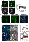
Figure 1.. TRPV4 is functionally expressed by enteric glia, macrophages, and endothelial cells of the mouse colon.
A. Enteric glia (arrows) and mMac (arrowheads) in the myenteric region exhibited increased [Ca2+]i in response to the TRPV4 agonist GSK101 (100 nM). Immunostaining confirmed the identity of glial cells (Sox10+: cyan, GFAP: green), mMac (white), and neurons (Hu+: magenta). Scale = 50 μm. B. Responses to GSK101 were also detected in glial cells (arrows) and macrophages (arrowheads) associated with submucosal ganglia. Scale = 50 μm. C. GSK101 evoked sustained Ca2+ signals in vascular (arrows, CD31+: white) and lymphatic (arrowheads, LYVE1+: green) endothelial cells. Each trace represents the response by an individual cell. Scale = 100 μm.
Figure 2.
Figure 2.. TRPV4 is functionally expressed by enteric glia and macrophages in the human colon.
A. GSK101 (100 nM) induced sustained Ca2+ responses in submucosal glia (arrowheads; S100B+: magenta) of the human colon. B. Equivalent responses were detected in the myenteric region. Insets i & ii show responses by myenteric glia (red arrows) and macrophages (magenta arrowheads; scale: 20 μm). Enteric glia were identified as non-neuronal cells (DAPI: blue) that were closely associated with neurons (Hu: green) within the ganglia. Macrophages were defined as IBA1+ cells (white). The grayscale images highlight responsive cells in white against a dark background (image normalized to baseline, F/F0). Scale = 50 μm.
Figure 3.
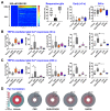
Figure 3.. TRPV4-mediated activation of the myenteric glial network of the mouse colon is mediated by gap junctions, purinergic activity, and release of Ca2+ from intracellular stores.
A. Heatmap demonstrating the oscillatory nature and varying kinetics of Ca2+ responses by glia to TRPV4 activation (100 nM GSK101, white dotted line). Responses by mMac to the same stimulus were sustained. A distinct subset of the GSK101-responsive glial population was identified that exhibited rapid (‘early’) elevations in Ca2+ within 5s of GSK101 application. The mechanism of GSK101-induced Ca2+ activity within the glial network was investigated by pharmacological inhibition of gap junctions (50 μM carbenoxolone: CBX), purinoceptors (50 μM suramin (SUR) + 50 μM PPADS), connexin43 hemichannels (50 μM GAP26), and depletion of intracellular Ca2+ from ER stores (30 μM cyclopiazonic acid: CPA). None of the pharmacological treatments significantly affected early responding glial populations (<5 s). However, there was a significant increase in the percentage of responding glia following CPA pre-treatment when assessed at 240 s. The kinetics of the Ca2+ responses were compared at t= 30 s (B) and 240 s (C). CBX pre-treatment resulted in a significant increase in AUC, halfwidth and rise-time (10-90%) for cells responding both within 30 s and 240 s post GSK101 addition, compared to vehicle. In the presence of CPA, there was an increase in rise-time for cells responding at both time points. (Vehicle: 1045 cells, N=9; CBX: 936 cells, N=8; CPA: 917 cells, N=7; Gap26: 503 cells, N=4; SUR+PPADS: 704 cells, N=6). (D) Pair correlation function (PCF) analysis of responses at t= 30 s for cells separated by 115 μm revealed smaller PCF values across all treatments compared to vehicle (PCF: Veh= 1.54, CBX= 0.93, SUR+PPADS= 1.02, CPA= 1.03, Gap26= 1.17). This indicates that the correlated glial activity was dependent on gap junctions, purinergic activity and intracellular Ca2+ stores. Data were analyzed by one-way ANOVA with Dunnett’s post hoc test, * P<0.05, ** P<0.01, **** P<0.0001. Data are expressed as mean ± 95% C.I. The horizontal line in each graph represents the mean, and whiskers denote the minimum and maximum values.
Figure 4.
Figure 4.. TRPV4-mediated glial activity is sensitized in acute TNBS-induced colitis and is associated with a reduction in correlated Ca2+ activity.
A. The percentage of GSK101 (100 nM)-responsive glia was increased in acutely inflamed tissues at both the early and late time points. B. GSK101-induced Ca2+ kinetics were not affected when assessed at 30 s post drug addition. However, there was an increase in the average number of peaks at 240 s (C). D. TRPV4-mediated Ca2+ responses in the glial network were less coordinated for cells within a 115 μm distance in inflamed tissues (PCF= 1.05) compared to the healthy control (PCF= 1.44). (Vehicle: 1260 cells, N=14; TNBS: 598 cells, N=7; one-way ANOVA with Dunnett’s post hoc test, * P<0.05, ** P<0.01. Data are expressed as mean ± 95% C.I. The horizontal line in the graph represents the mean, and whiskers denote the minimum and maximum values.
Figure 5.
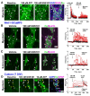
Figure 5.. Enteric neurons of the mouse colon do not express functional TRPV4.
A. Wholemount preparation loaded with Fluo8-AM dye. Myenteric neurons (asterisks; Hu+: green) responded to ATP (100 μM) but were unresponsive to GSK101 (100 nM). In contrast, enteric glia (arrowheads; GFAP+: magenta) responded to both agonists. B-C. Wholemount preparation of the Wnt1-GCaMP3 mouse colon. GSK101 evoked Ca2+ responses only in enteric glia (arrowheads; Sox10+: magenta) and not enteric neurons (asterisks; Hu+: green) (representative examples from n=5). D. Cultured enteric glia (yellow arrowheads; GFAP+: magenta), but not myenteric neurons (white arrowheads; Hu+: green), exhibited increased [Ca2+]i in response to GSK101. Both neurons and glia responded to subsequent exposure to ATP (10 μM). In contrast to observations from tissue-based imaging, GSK101-mediated [Ca2+]i responses by glia were sustained (n=3 independent cultures). Scale = 40 μm.
Figure 6.
Figure 6.. TRPV4 activation does not affect contractility of the isolated mouse colon.
A. Representative trace demonstrating no effect of GSK101 (100 nM) on circular muscle tension relative to vehicle (DMSO). Tissues contracted in response to the positive control carbachol (CCh, 10 μM; ***p<0.001, n=9 mice, one-way ANOVA, Tukey’s post-hoc test). B. TRPV4 activation (GSK101, 100 nM) had no effect on the mean amplitude of electrically evoked neurogenic contractions relative to DMSO control. A representative trace of responses to EFS (•) recorded at baseline and following subsequent addition of DMSO and GSK101 (100 nM) is shown (p=0.084, n=6, paired t-test). C. Representative DMaps showing propagating contractions (vertical white lines) under threshold intraluminal pressure in the presence of vehicle (DMSO, left) then GSK101 (100 nM, right). D. (a) The interval between CMCs and mean colon diameter (b) was unaffected by pharmacological activation (left: 100nM GSK101) or inhibition (middle: 1μM GSK219) of TRPV4, or by TRPV4 knockout (right). (c) CMC velocity was not changed in response to GSK101 (left) or TRPV4 deletion (right) but was significantly enhanced in the presence of GSK219 (middle). Points represent mean values per preparation, n=9-20 mice per group. Data were compared by paired (GSK101, GSK219) or unpaired (TRPV4−/−) two-tailed t-tests. E. Posterior distribution graphs demonstrate the probability of a contraction occurring at different colon diameters under control or GSK101-treated conditions. Colored dots represent data from independent experiments (n=34 and 24 mice). No difference in mean gut diameter was detected between control and GSK101-treated preparations (ratio of the mean diameter= 1.03). F. Posterior distribution graphs summarizing the mean rate of contraction under control and GSK101-treated conditions (n=34 and 24 mice). There was no difference in the rate of contractions between the two groups (ratio of the mean= 1.04).

Similar articles

References

    1. Spencer NJ, Zagorodnyuk V, Brookes SJ, Hibberd T. Spinal afferent nerve endings in visceral organs: recent advances. Am J Physiol Gastrointest Liver Physiol 2016;311:G1056–G63. - PubMed
    1. Treichel AJ, Finholm I, Knutson KR, Alcaino C, Whiteman ST, Brown MR, et al. Specialized Mechanosensory Epithelial Cells in Mouse Gut Intrinsic Tactile Sensitivity. Gastroenterology 2022;162:535–47 e13. - PMC - PubMed
    1. Servin-Vences MR, Lam RM, Koolen A, Wang Y, Saade DN, Loud M, et al. PIEZO2 in somatosensory neurons controls gastrointestinal transit. Cell 2023;186:3386–99 e15. - PMC - PubMed
    1. Brierley SM, Page AJ, Hughes PA, Adam B, Liebregts T, Cooper NJ, et al. Selective role for TRPV4 ion channels in visceral sensory pathways. Gastroenterology 2008;134:2059–69. - PMC - PubMed
    1. Cenac N, Altier C, Chapman K, Liedtke W, Zamponi G, Vergnolle N. Transient receptor potential vanilloid-4 has a major role in visceral hypersensitivity symptoms. Gastroenterology 2008;135:937–46, 46 e1-2. - PubMed

Publication types