[Preprint]. 2024 Jan 9:2024.01.08.574768.
doi: 10.1101/2024.01.08.574768.

Primary oocytes with cellular senescence features are involved in ovarian aging in mice

Affiliations

Primary oocytes with cellular senescence features are involved in ovarian aging in mice

Hao Yan et al. bioRxiv. .

Update in

Abstract

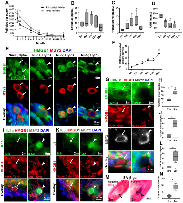
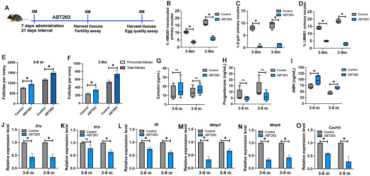
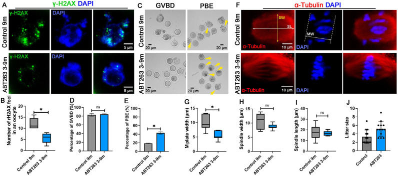
In mammalian females, quiescent primordial follicles serve as the ovarian reserve and sustain normal ovarian function and egg production via folliculogenesis. The loss of primordial follicles causes ovarian aging. Cellular senescence, characterized by cell cycle arrest and production of the senescence-associated secretory phenotype (SASP), is associated with tissue aging. In the present study, we report that some quiescent primary oocytes in primordial follicles become senescent in adult mouse ovaries. The senescent primary oocytes share senescence markers characterized in senescent somatic cells. The senescent primary oocytes were observed in young adult mouse ovaries, remained at approximately 15% of the total primary oocytes during ovarian aging from 6 months to 12 months, and accumulated in aged ovaries. Administration of a senolytic drug ABT263 to 3-month-old mice reduced the percentage of senescent primary oocytes and the transcription of the SASP cytokines in the ovary. In addition, led to increased numbers of primordial and total follicles and a higher rate of oocyte maturation and female fertility. Our study provides experimental evidence that primary oocytes, a germline cell type that is arrested in meiosis, become senescent in adult mouse ovaries and that senescent cell clearance reduced primordial follicle loss and mitigated ovarian aging phenotypes.


Figures

Figure 1.
Figure 1.. Adult mouse ovaries contained primary oocytes that were positive for makers of cellular senescence during aging.
(A) Numbers of primordial follicles (black line) and total follicles (grey line) in the ovary declined significantly during aging. (B-D) Changes in the levels of estradiol (B), progesterone (C), and AMH (D) in the serum during mouse aging. (E) Primary oocytes stained HMGB1 positive in the nucleus and negative in the cytoplasm (arrows, nuc+;cyto−) found in 2-month (2m) ovaries; and primary oocytes with translocated HMGB1 staining: HMGB1 positive in both the nucleus and cytoplasm (arrowheads, nuc+;cyto+), HMGB1 negative in the nucleus and positive in the cytoplasm (arrowheads, nuc−;cyto+), and HMGB1 negative in both the nucleus and cytoplasm ( arrowheads, nuc−;cyto−). (F) The percentage of primary oocytes with translocated HMGB1 increased during mouse aging. (G) The primary oocyte that was only nucleus-positive for HMGB1 stained positive for nuclear envelope protein lamin b1 (arrows, LMNB1+); the primary oocyte had translocated HMGB1 staining (Nuc−; Cyto−) stained negative for lamin b1 (arrowheads, LMNB1−). (H) The percentage of primary oocytes had LMNB1 negative staining in 2-month and 9-month ovaries. (I) The primary oocyte that was only nucleus-positive for HMGB1 stained negative for IL1a (IL1a-; arrows); the primary oocyte had translocated HMGB1 staining (Nuc−; Cyto−) stained positive for IL1a (IL1a+; arrowheads). (J) The percentage of primary oocytes had IL1a positive staining in 2-month and 9-month ovaries. (K) The primary oocyte that was nucleus-positive for HMGB1 stained negative for IL6 (IL6-; arrows); the primary oocyte with translocated HMGB1 staining (Nuc−; Cyto−) stained positive for IL6 (IL6+; arrowheads). (L) The percentage of primary oocytes had IL6-positive staining in 2-month and 9-month ovaries. (M) Primary oocyte with SA-β-gal negative staining (arrow) and positive staining (arrowhead). (N) The percentage of primary oocytes had SA-β-gal positive staining in 2-month and 9-month ovaries. Data in the graph are presented as mean ± SD. * represents significant difference.
Figure 2.
Figure 2.. Senolytic drug ABT263 treatment mitigated ovarian aging phenotypes.
(A) Schematic timeline of ABT263 treatment. (B-D) Percentages of HMGB1 translocated primary oocytes (B), β-gal-positive primary oocytes (C), and LMNB1-negative primary oocytes (D) in control and ABT263-treated ovaries at 6 months and 9 months. (E-F) Numbers of primordial follicles and total follicles in control and ABT263-treated ovaries at 6 months (E) and 9 months (F). (G-I) Levels of estradiol (G), progesterone (H), and AMH (I) in the serum of control mice and ABT263-treated mice at 6 months and 9 months. (J-O) Relative expression of mRNAs of Il1a (J), Il1b (K), Il6 (L), Mmp3 (M), Mmp9 (N), and Cxcl10 (O) in control and ABT263-treated ovaries at 6 months and 9 months. Data in the graph are presented as mean ± SD. * represents significant difference and ns represents no significant difference.
Figure 3.
Figure 3.. Effect of ABT263 treatment on oocyte maturation and fertility.
(A) Oocytes from 9-month-old control mice (control 9m) and ABT263-treated mice (ABT263 3-9m) were stained with an antibody to γ-H2AX to detect DNA damage. DNA is revealed by DAPI staining. (B) Number of γ-H2AX-positive foci in the oocytes isolated from control ovaries and ABT263-treated ovaries at 9 months. (C) Brightfield microscopic images showing that oocytes isolated from control ovaries and ABT263-treated ovaries at 9 months underwent germinal vesicle breakdown (GVBD), and more oocytes isolated from ABT263-treated ovaries completed first polar body extrusion (PBE, arrows). (D, E) Percentages of oocytes underwent GVBD (D) and PBD (E). (F) Examples of meiotic spindles observed in oocytes isolated from control ovaries and ABT263-treated ovaries at 9 months. (G-I) The width of the metaphase I plate (MW) (G), the width of the meiotic spindle (SW) (H), and the length of the meiotic spindle (SL) (I) in the oocytes isolated from control ovaries and ABT263-treated ovaries at 9 months. (J) Numbers of pups from each litter of control mice and ABT263-treated mice during fertility assay. Data in the graph are presented as mean ± SD. * represents significant difference and ns represents no significant difference.
Figure 4.
Figure 4.. Comparative transcriptome profiling of the ovaries during aging and between the ovaries of ABT263-treated mice and control mice.
(A-D) Gene ontology analysis of biological pathways (BP) that were significantly upregulated in the ovaries of 6-month-old mice (A) and 9-month-old mice (B) compared with 3-month-old mice; and the BPs that were significantly downregulated in the ovaries of 6-month-old mice (C) and 9-month-old mice (D) compared with 3-month-old mice. (E, F) BPs that were significantly upregulated (E) or downregulated (F) in 6-month-old ovaries of ABT263-treated mice compared with that of control mice. (G, H) BPs that were significantly upregulated (G) or downregulated (H) in 9-month-old ovaries of ABT263-treated mice compared with that of control mice. (I) Heatmap showing fold change in mRNA expression of SASP cytokines and proteins in the ovaries during aging and between the ovaries of ABT263-treated mice and control mice.

References

    1. Pelosi E., Forabosco A., and Schlessinger D., Genetics of the ovarian reserve. Front Genet, 2015. 6: p. 308. - PMC - PubMed
    1. Pepling M.E., Follicular assembly: mechanisms of action. Reproduction, 2012. 143(2): p. 139–49. - PubMed
    1. Gougeon A., Dynamics of follicular growth in the human: a model from preliminary results. Hum Reprod, 1986. 1(2): p. 81–7. - PubMed
    1. Lopez J., et al., The Aging Ovary and the Tales Learned Since Fetal Development. Sex Dev, 2023. 17(2-3): p. 156–168. - PMC - PubMed
    1. Broekmans F.J., Soules M.R., and Fauser B.C., Ovarian aging: mechanisms and clinical consequences. Endocr Rev, 2009. 30(5): p. 465–93. - PubMed

Publication types