[Preprint]. 2024 Jan 3:2024.01.02.573945.
doi: 10.1101/2024.01.02.573945.

A "double-edged" role for type-5 metabotropic glutamate receptors in pain disclosed by light-sensitive drugs

Affiliations

A "double-edged" role for type-5 metabotropic glutamate receptors in pain disclosed by light-sensitive drugs

Serena Notartomaso et al. bioRxiv. .

Update in

Abstract

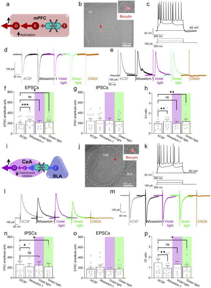
Knowing the site of drug action is important to optimize effectiveness and address any side effects. We used light-sensitive drugs to identify the brain region-specific role of mGlu5 metabotropic glutamate receptors in the control of pain. Optical activation of systemic JF-NP-26, a caged, normally inactive, negative allosteric modulator (NAM) of mGlu5 receptors, in cingulate, prelimbic and infralimbic cortices and thalamus inhibited neuropathic pain hypersensitivity. Systemic treatment of alloswitch-1, an intrinsically active mGlu5 receptor NAM, caused analgesia, and the effect was reversed by light-induced drug inactivation in in the prelimbic and infralimbic cortices, and thalamus. This demonstrates that mGlu5 receptor blockade in the medial prefrontal cortex and thalamus is both sufficient and necessary for the analgesic activity of mGlu5 receptor antagonists. Surprisingly, when light was delivered in the basolateral amygdala, local activation of systemic JF-NP-26 reduced pain thresholds, whereas inactivation of alloswitch-1 enhanced analgesia. Electrophysiological analysis showed that alloswitch-1 increased excitatory synaptic responses in prelimbic pyramidal neurons evoked by stimulation of BLA input, and decreased feedforward inhibition of amygdala output neurons by BLA. Both effects were reversed by optical silencing and reinstated by optical reactivation of alloswitch-1. These findings demonstrate for the first time that the action of mGlu5 receptors in the pain neuraxis is not homogenous, and suggest that blockade of mGlu5 receptors in the BLA may limit the overall analgesic activity of mGlu5 receptor antagonists. This could explain the suboptimal effect of mGlu5 NAMs on pain in human studies and validate photopharmacology as an important tool to determine ideal target sites for systemic drugs.


Conflict of interest statement

Competing interests: The authors declare that they have no competing interests.

Figures

Fig. 1.
Fig. 1.. Neuropathic pain model and mGlu5 receptor signaling in different brain regions.
(a) Experimental protocol. (b) Mechanical (hyper-)sensitivity on the left hindpaw measured with von Frey filaments was reduced in CCI mice (n = 10) versus sham-operated mice (n = 10) 16 days after nerve ligation. Bar histograms show means ± SEM. (c-g) mGlu5 receptor mediated PI hydrolysis in different brain regions of sham and CCI mice. Increased InsP levels were observed in infralimbic (c), prelimbic (p=0.051) (d) and cingulate (e) cortices and amygdala (f), but not thalamus (g), of neuropathic mice (CCI) after stimulation with a positive allosteric modulator (PAM, VU0360172, 10 mg/kg i.p.) compared to vehicle. Enhanced InsP concentration was detected in the thalamus, but not in the prefrontal cortical regions and amygdala, of sham animals treated with VU0360172 compared to vehicle. Bar histograms on the right in c-g show percent increase above vehicle in sham and CCI mice. Bar histograms show means ± SEM of 10 (b), 3–5 (c), 4–5 (d), 4–5 (e), 5–8 (f), 10 (g) mice per group. Two-way ANOVA: (b) sham vs CCI, F(1,36)=16.04; p=0.0003; ipsi vs contra, F(1,36)=7.304; p=0.0104; interaction F(1,36)=5.062, p=0.0307; (c) sham vs CCI, F(1,11)=0.5569; p=0.4712; vehicle vs VU0360172, F(1,11)=5.741, p=0.0355; interaction (F1,11)=0.8168, p=0.3855; (d) sham vs CCI, F(1,15)=1.075; p=0.3163; vehicle vs VU0360172, F(1,15)=6.452, p=0.0226; interaction (F1,15)=0.7034, p=0.4148; (e) sham vs CCI, F(1,13)=3.951; p=0.0683; vehicle vs VU0360172, F(1,13)=11.21, p=0.0052; interaction (F1,13)=0.1553, p=0.6999; (f) sham vs CCI, F(1,22)=0.01997, p=0.8889; vehicle vs VU0360172, F(1,22)=8.888, p=0.0069; interaction (F1,22)=6.099, p=0.0218; (g) sham vs CCI, F(1,36)=0.4861, p=0.4902; vehicle vs VU0360172, F(1,36)=4.557, p=0.0397; interaction (F1,36)=2.313, p=0.1370. *p<0.05, **p<0.01; ***p<0.001, Bonferroni’s multiple comparison post hoc test.
Fig. 2.
Fig. 2.. Behavioral effects of light-induced manipulations of mGlu5 receptors in different brain regions in CCI mice.
Schematic representation of sites of stereotaxic implantation of LED optical fibers in different brain regions. (b) Experimental protocol for optical modulation. (c) Optical activation (blue-violet light, 405 nm) of mGlu5 NAM JF-NP-26 and optical inactivation (blue-violet light, 405 nm) and reactivation (green light, 520 nm) of mGlu5 NAM alloswitch-1. (d-i) Optical activation (blue-violet light, 405 nm) of mGlu5 NAM JF-NP-26 (10 mg/kg, i.p.) in contralateral (to the side of CCI) infralimbic cortex (d), prelimbic cortex (e), anterior cingulate cortex (f) and thalamus (g) caused a significant increase in mechanical thresholds, whereas JF-NP-26 activation in the amygdala (h) caused hyperalgesia, in neuropathic mice 16 days after CCI induction (i). (j-o) Systemic application of mGlu5 NAM alloswitch-1 (10 mg/kg, i.p.) caused antinociception in CCI mice, compared to vehicle. Optical inactivation (blue-violet light, 405 nm) of alloswitch-1 in the contralateral (to the side of CCI) infralimbic cortex (j), prelimbic cortex (k), or thalamus (n) reversed the antinociceptive effects, while light-induced (green light, 520 nm) reactivation of alloswitch-1 in those brain regions reinstated analgesia. No significant behavioral changes were observed with optical manipulations in anterior cingulate cortex (l). In contrast, optical inactivation of alloswitch-1 in the amygdala with blue-violet light further increased mechanical thresholds (enhancement of antinociception), and this effect was reversed by reactivation of alloswitch-1 with green light (m). Bar histograms show mean ± SEM of 8 (d), 7 (e), 11 (f and g), 6 (h), 7 (j), 9 (k), 8 (l), 6 (m) and 5 (n) mice per group. *p<0.05, ***p<0.001, paired student’s t-test compared to JF-NP-26 without light activation. (d) t=2.487, p=0.0418; (e) t=3.243, p=0.0176; (f) t=2.390, p=0.0379; (g) t=3.025, p=0.0128; (h) t=9.062, p=0.0003. One-way repeated measures ANOVA; (j) F(3,18)=19.25, p<0.0001; (k) F(3,24)=20.12, p<0.0001; (l) F(3,21)=3.813, p=0.0251; (m) F(3,15)=24.66, p<0.0001; (n) F(3,12)=5.463, p=0.0133. Bonferroni’s multiple comparisons post hoc test. *p<0.05, **p<0.01, ***p<0.001.
Fig. 3.
Fig. 3.. Electrophysiological effects of light-induced off/on-switch alloswitch-1 on prelimbic pyramidal neurons and amygdala feed-forward inhibition in neuropathic pain.
(a) Prelimbic circuitry to explain mGlu5 receptor action. Inhibitory (I) interneuron projects onto excitatory (E) inputs to pyramidal output neurons (O). (b, c) Whole-cell patch-clamp electrophysiological recordings were performed visually identified layer 5 pyramidal neurons in brain slices obtained from CCI mice 16 days after induction. Traces recorded in an individual neuron show typical regular action potential firing pattern in response to intracellular depolarizing current injections. (d, e) Excitatory postsynaptic currents (EPSCs, recorded at −70 mV; d) and inhibitory postsynaptic currents (IPSCs, recorded at 0 mV; e) were evoked by focal electrical stimulation of fibers of passage in the infralimbic cortex. EPSCs and IPSCs were blocked by a glutamate receptor antagonist (CNQX, 20 μM) confirming glutamatergic EPSCs and glutamate-driven IPSCs. Synaptic responses were evaluated before (ASCF) and during alloswitch-1 bath application, and under blue-violet and green light illumination. (f) Alloswitch-1 (100 nM by superfusion) enhanced the peak amplitude of EPSCs. (g) EPSCs were not different from baseline with blue-violet light-induced inactivation of alloswitch-1 (5 min, 0.5 Hz, 500 ms) while subsequent reactivation under green light illumination (5 min, 0.5 Hz, 500 ms) significantly increased EPSCs compared to pre-drug values. (g) No significant changes were observed on the IPSCs. (h) Changes in excitatory/inhibitory (E/I) ratio. (i) Amygdala circuitry to explain mGlu5 receptor action. Excitatory (E) input from basolateral to central nucleus engages inhibitory (I) signaling for feedforward inhibition of output (O) neurons. (j, k) Whole-cell patch-clamp electrophysiological recordings were performed from amygdala neurons (latero-capsular division, CeLC) of brain slices obtained from CCI mice 16 days after induction. (l, m) Glutamate-driven IPSCs (recorded at 0 mV; l) and monosynaptic EPSCs (recorded at −70 mV; IPSCs and EPSCs were blocked by CNQX, 20 μM, confirming glutamate-driven IPSCs (feedforward inhibition) and glutamatergic EPSCs; m) were evoked in CeLC neurons by focal electrical stimulation in the BLA. Synaptic responses were evaluated before (ASCF) and during alloswitch-1 bath application, and under blue-violet and green light illumination. (n) Alloswitch-1 (100 nM) significantly decreased the peak amplitude of the IPSCs; IPSCs were not significantly different from baseline with blue-violet light-induced inactivation of alloswitch-1 (5 min, 0.5 Hz, 500 ms), whereas subsequent drug reactivation under green light illumination (5 min, 0.5 Hz, 500 ms) significantly decreased IPSCs compared to pre-drug values. (o) No significant changes were observed on the EPSCs. (p) Changes in inhibitory/excitatory (I/E) ratio. The data suggest that mGlu5 blockade in the CeA reduced BLA-driven feed-forward inhibition onto the CeLC neurons resulting in behavioral hypersensitivity. Bar histograms show mean ± SEM of n=10 in 7 mice (f, g, h) and n=12 in 8 mice (n, o, p) neurons. One-way ANOVA repeated measures: (f) F(3.27)=6.323, p=0.0022; (g) F(3,27)=0.9374, p=0.4362; (h) F(3,27)=5.499, p=0.0044; (n) F(3,33)=4.079, p=0.0144; (o) F(3,33)=1.779, p=0.1704; (p) F(3,33)=5.581, p=0.0033. Dunnett’s multiple comparisons post hoc test. *p<0.05; **p<0.01 compared to pre-drug.
Fig. 4.
Fig. 4.. Behavioral and neuronal effects of brain region-specific modulation of mGlu5 receptors.
(a) Neuropathic pain behavior was inhibited by mGlu5 receptor blockade in prefrontal cortex and thalamus with opposite effects in the amygdala. (b) Hypothesized neural circuitry based on electrophysiological data. mGlu receptor blockade decreases feedforward inhibition from basolateral to central nucleus to increase amygdala output but has no effect on prefrontal cortex. mGlu receptor blockade in prelimbic cortex decreases inhibition of excitatory synaptic drive onto pyramidal output neurons and decrease activation of pronociceptive RVM ON-cells. mPFC = medial prefrontal cortex; VPL = ventral posterolateral nucleus of the thalamus; BLA = basolateral amygdala; CeA = central nucleus of amygdala; RVM = rostral ventromedial medulla; O = output neuron; E = excitatory neuron; I = inhibitory neuron.

References

    1. Rodriguez E. A., Campbell R. E., Lin J. Y., Lin M. Z., Miyawaki A., Palmer A. E., Shu X., Zhang J., Tsien R. Y., The Growing and Glowing Toolbox of Fluorescent and Photoactive Proteins. Trends Biochem Sci 42, 111–129 (2017). - PMC - PubMed
    1. Wu Y., Shroff H., Multiscale fluorescence imaging of living samples. Histochem Cell Biol 158, 301–323 (2022). - PMC - PubMed
    1. Szobota S., Isacoff E. Y., Optical control of neuronal activity. Annu Rev Biophys 39, 329–348 (2010). - PMC - PubMed
    1. Paoletti P., Ellis-Davies G. C. R., Mourot A., Optical control of neuronal ion channels and receptors. Nat Rev Neurosci 20, 514–532 (2019). - PMC - PubMed
    1. Hull K., Morstein J., Trauner D., In Vivo Photopharmacology. Chem Rev 118, 10710–10747 (2018). - PubMed

Publication types