[Preprint]. 2024 Jan 11:2024.01.10.575114.
doi: 10.1101/2024.01.10.575114.

SARS-CoV-2 papain-like protease activates nociceptors to drive sneeze and pain

Affiliations

SARS-CoV-2 papain-like protease activates nociceptors to drive sneeze and pain

Sonali S Mali et al. bioRxiv. .

Abstract

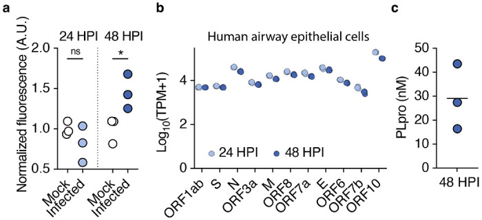
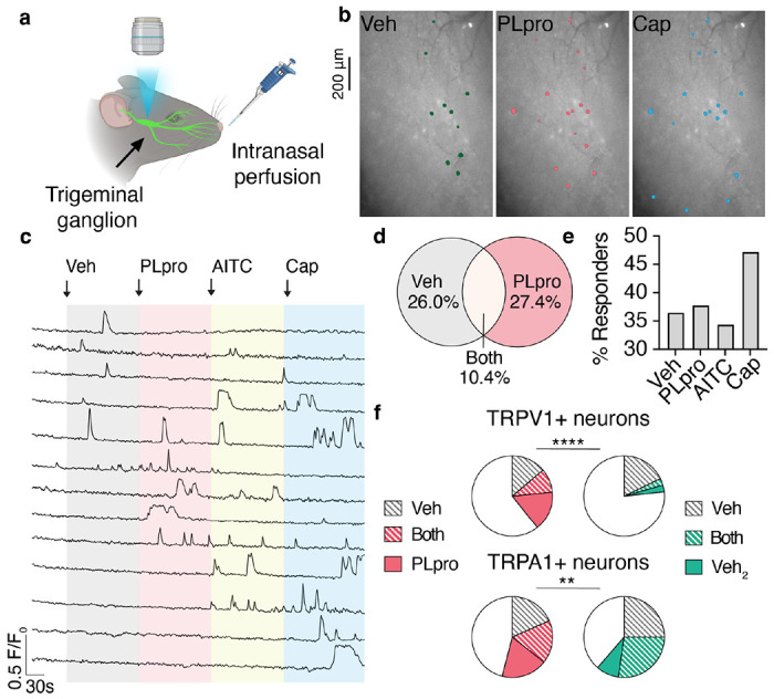
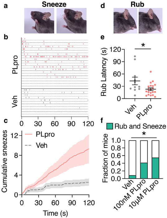
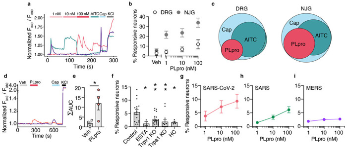
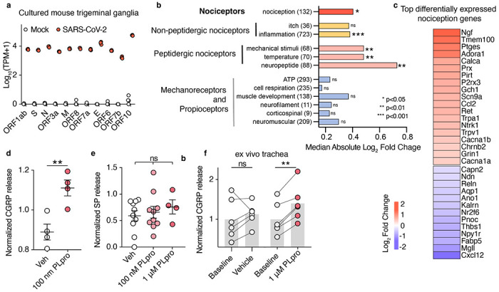
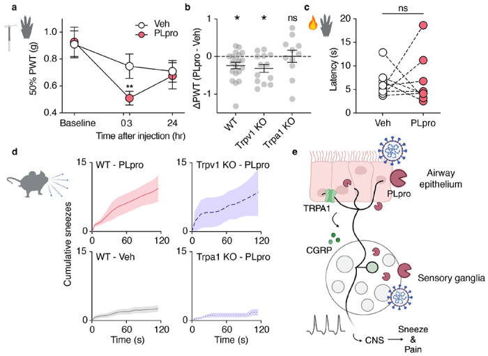
SARS-CoV-2, the virus responsible for COVID-19, triggers symptoms such as sneezing, aches and pain.1 These symptoms are mediated by a subset of sensory neurons, known as nociceptors, that detect noxious stimuli, densely innervate the airway epithelium, and interact with airway resident epithelial and immune cells.2-6 However, the mechanisms by which viral infection activates these neurons to trigger pain and airway reflexes are unknown. Here, we show that the coronavirus papain-like protease (PLpro) directly activates airway-innervating trigeminal and vagal nociceptors in mice and human iPSC-derived nociceptors. PLpro elicits sneezing and acute pain in mice and triggers the release of neuropeptide calcitonin gene-related peptide (CGRP) from airway afferents. We find that PLpro-induced sneeze and pain requires the host TRPA1 ion channel that has been previously demonstrated to mediate pain, cough, and airway inflammation.7-9 Our findings are the first demonstration of a viral product that directly activates sensory neurons to trigger pain and airway reflexes and highlight a new role for PLpro and nociceptors in COVID-19.


Figures

Figure 1.
Figure 1.. SARS-CoV-2 PLpro is released from infected airway epithelial cells.
a, PLpro proteolytic activity from the supernatant of SARS-CoV-2 or mock infected Calu-3 cells 24 and 48 HPI, measured by the fluorescence of cleaved substrate, normalized across samples from the same day. One-way ANOVA: p=0.014, F(3,8)=6.790; Holm-šídák’s multiple comparisons, pmock vs. infected 24h =0.244, pmock vs. infected 48h =0.030, n=3 wells per group. b, SARS-CoV-2 transcripts from Calu-3 cells infected with SARS-CoV-2 (multiplicity of infection = 1) for 24 and 48 hours post infection (HPI), transcripts per million (TPM), n=3 wells. c, Interpolated concentration of PLpro in the supernatant of Calu-3 cells 48 hours post infection, line represents mean.
Figure 2.
Figure 2.. PLpro activates upper airway-innervating neurons in vivo.
a, Schematic of in vivo imaging preparation. b, Image of trigeminal ganglion overlaid with the regions of interest of neurons that respond to intranasal perfusion (10 μL) of vehicle (Veh), 10 μM PLpro, and 100 μM capsaicin (Cap). c, Representative calcium transients in response to intranasal perfusion of Veh, 10 μM PLpro, 1 mM AITC, 100 μM Cap. d, Venn diagram of neurons responsive to vehicle (112/307) and PLpro (116/307), n=307 neurons from 6 mice. PLpro activates a subset of vehicle-sensitive neurons (32/112). e, Percent of recorded neurons that respond to each stimulus. f, Vehicle and PLpro activate a subset of TRPV1+ (capsaicin-responsive) neurons and TRPA1+ (AITC-responsive) neurons. Top Left, percent of TRPV1+ neurons responsive to vehicle only: 13.8%, both vehicle and PLpro: 9.7%, PLpro only: 15.9%, n=145 neurons from 6 mice; Top Right, percent of TRPV1+ neurons responsive to the first delivery of vehicle only: 17.8%, both first and second (Veh2) deliveries of vehicle: 2.7%, second delivery of vehicle only: 2.7%, n=73 neurons from 3 mice, Chi-square test χ2 = 112.5, df = 3, p<0.0001. Bottom Left, percent of TRPA1+ neurons responsive to vehicle only: 18.4%, both vehicle and PLpro: 17.2%, PLpro only: 18.4%, n=87 neurons from 4 mice; Bottom Right, percent of TRPA1+ neurons responsive to the first delivery of vehicle only: 25.0%, both deliveries of vehicle: 27.3%, second delivery of vehicle only: 9.1%, n=44 neurons from 3 mice, Chi-square test χ2 = 14.2, df = 3, p = 0.003.
Figure 3.
Figure 3.. PLpro elicits sneezes and nose rubs in mice.
a, Sequential images of a sneeze following intranasal treatment of 10 μM PLpro (10 μL). b, Raster plot of sneezes from individual mice treated with 10 μM PLpro or vehicle identified from audio recordings. c, Average cumulative sneeze count of 10 μM PLpro and vehicle treated mice over 2 minutes d, Sequential images of a nose rub following intranasal treatment e, 10 μM PLpro treated mice display a shorter latency to the first nose rub than vehicle treated mice, t-test: t=2.469, df=29, p=0.020. f, Fraction of mice that elicited a rub and sneeze within the first 30 seconds after treatment. (Vehicle: 9.0%, PLpro (100 nM): 41.7%, PLpro (10 μM): 55.0%). Fisher’s exact test (pvehicle vs. 100nM PLpro =0.252, pvehicle vs. 10μM PLpro =0.020). For all data in this figure, vehicle (n=11), 100 nM PLpro (n=12), and 10 μM PLpro (n=20). Error bars and shading represent the mean ± standard error of the mean (SEM), n = biological replicates (animals).
Figure 4.
Figure 4.. PLpro directly activates nociceptors.
a, Representative calcium transients from PLpro responders from cultured neonatal mouse trigeminal ganglia (TG). b, PLpro activates subsets of neurons from adult mouse dorsal root ganglia (DRG; Veh: 2.9%, 1 nM: 4.2%, 10 nM: 6.8%, 100 nM: 11.9%, n=5) and adult mouse nodose and jugular ganglia (NJG; Veh: 5.5%, 1 nM: 21.7%, 10 nM: 23.6%, 100 nM: 33.9%, n=8). c, Venn diagram of PLpro, AITC, and Cap-responsive neurons in DRG and NJG. Of the neurons that respond to these 3 stimuli, Left, in the DRG, 20% responded to PLpro, an additional 48% responded to AITC, and 32% responded to Cap alone. Right, in the NJG, 38% responded to PLpro, an additional 34% responded to AITC, and 28% responded to Cap alone. d, Representative calcium transients evoked by PLpro and capsaicin (Cap) in human iPSC-derived neurons. e, PLpro induces significantly more calcium influx (area under the curve, AUC) than vehicle, t-test: t=2.953, df=6, p=0.026, n=4 wells each. f, Percent of neonatal mouse TG neurons that respond to 250nM PLpro in control neurons from wild-type C57BL/6N mice in physiological extracellular calcium (2 mM Ca2+, 5.5%, n=17) is greater than in the absence of extracellular calcium (EGTA, 1.2%, n=3) and in neurons from Trpv1 KO (2.4%, n=27) and Trpa1 KO (1.9%, n=12) mice, and in wild-type mice in the presence of TRPA1 antagonist (HC, 1.7%, n=4). One-way ANOVA: p=0.003, F(4,58)=2.803; Holm-šídák’s multiple comparisons, pControl vs. EGTA =0.035, pControl vs. Trpv1 KO =0.004, pControl vs. Trpal KO =0.004, pControl vs. EGTA =0.035. g-i, SARS-CoV-2 and SARS, but not MERS PLpro activate subsets of neonatal mouse TG neurons in a dose-dependent manner. g, SARS-CoV-2 PLpro: 1nM: 3.8%, 10 nM: 7.2%, 100 nM: 9.3%, n=5. h, SARS PLpro: 1 nM: 1.4%, 10 nM: 3.4%, 100nM: 6.1%, n=5. i, MERS PLpro: 1nM: 1.8%, 10nM: 2.5%, 100nM: 2.7%, n=6. *p <0.05, **p<0.01. Error bars represent the mean ± SEM, n indicates replicates (wells).
Figure 5.
Figure 5.. SARS-CoV-2 infection and PLpro activation share a key nociceptive signature.
a, SARS-CoV-2 viral transcript expression 48 hours after infection in cultured adult trigeminal ganglia, mock infected: n=4 wells, SARS-CoV-2 infected: n=3 wells. b, Infection alters gene sets associated with nociception but not mechanoreceptors and proprioceptors. The median absolute log2 fold change from each custom gene set was tested against 100,000 permutations of randomly selected gene sets of the same size, size of each gene set listed in parenthesis. c, Heatmap of the most highly differentially expressed nociception-associated transcripts (log2 fold change of infection vs mock). d, PLpro elicits CGRP release from cultured TG. t-test: t=3.928, df=6, p=0.008, n = 4 per group, biological replicates (animal) e, PLpro does not elicit the release of Substance P from cultured TG, One-way ANOVA: p=0.673, F(2,21)=0.215, vehicle (n=10), 100 nM PLpro (n=10), and 1 μM PLpro (n=4) replicates (wells from pooled animals) f, PLpro but not vehicle increases CGRP release from baseline measurements in an ex vivo trachea preparation. Baseline vs. vehicle: one sample t-test: t=0.909, df=5, p=0.405, baseline vs. 1μM PLpro: one sample t-test: t=6.80, df=5, p=0.001, n = 6, biological replicates (animal) per group. *p <0.05, **p<0.01, ***p<0.001. Bar represents mean and error bars represent the mean ± SEM.
Fig 6.
Fig 6.. SARS-CoV-2 PLpro-evoked sneeze and pain require TRPA1.
a, 50 μM PLpro induces a reduction in the paw withdrawal threshold (PWT) for force 3, but not 24 hours post injection in PLpro injected paws, but not vehicle injected paws compared to baseline. (24 hours pre-injection). Two-way ANOVA: ptime = 0.008, F(1.793, 64.56) = 5.477; Holm-šídák’s multiple comparisons, Veh: p-adjustedBaseline vs. 3 hours =0.361, p-adjustedBaseline vs. 24 hours =0.079; PLpro: p-adjustedBaseline vs. 3 hours =0.008, p-adjustedBaseline vs. 24 hours =0.117, n= 19. b, Wild-type C57BL/6J (WT) and Trpv1 KO mice, but not Trpa1 KO mice develop mechanical hypersensitivity 3 hours post injection. WT: one sample t-test: t=2.746, df=19, p=0.013, n = 11; Trpv1 KO: one sample t-test: t=3.001, df=20, p=0.011, n = 13; Trpa1 KO: one sample t-test: t=0.039, df=10, p=0.970, n = 11. c, 50 μM PLpro injection does not change the latency to respond to the radiant heat stimulation, paired t-test: t=0.233, df=7, p=0.822, n = 8. d, Intranasal treatment of 10 μM PLpro triggers more sneezing in Top Left, WT and Top Right, Trpv1 KO mice than Bottom Right, Trpa1 KO mice or Bottom Left, WT mice treated with vehicle. e, Proposed model: PLpro released from infected cells in the airway epithelium and in the sensory ganglia activate Trpa1-expressing neurons to drive sneeze, pain, and the release of CGRP into the airways. Error bars and shading represent the mean ± standard error of the mean (SEM), n = biological replicates (animals).

References

    1. Bliddal S. et al. Acute and persistent symptoms in non-hospitalized PCR-confirmed COVID-19 patients. Sci. Rep. 11, 13153 (2021). - PMC - PubMed
    1. Li F. et al. Sneezing reflex is mediated by a peptidergic pathway from nose to brainstem. Cell 184, 3762–3773.e10 (2021). - PMC - PubMed
    1. Finger T. E., St Jeor V. L., Kinnamon J. C. & Silver W. L. Ultrastructure of substance P- and CGRP-immunoreactive nerve fibers in the nasal epithelium of rodents. J. Comp. Neurol. 294, 293–305 (1990). - PubMed
    1. Baral P. et al. Nociceptor sensory neurons suppress neutrophil and γδ T cell responses in bacterial lung infections and lethal pneumonia. Nat. Med. 24, 417–426 (2018). - PMC - PubMed
    1. Pinho-Ribeiro F. A., Verri W. A. Jr & Chiu I. M. Nociceptor Sensory Neuron-Immune Interactions in Pain and Inflammation. Trends Immunol. 38, 5–19 (2017). - PMC - PubMed

Publication types