[Preprint]. 2024 Jan 11:2024.01.11.575259.
doi: 10.1101/2024.01.11.575259.

SAX-7/L1CAM acts with the adherens junction proteins MAGI-1, HMR-1/Cadherin, and AFD-1/Afadin to promote glial-mediated dendrite extension

Affiliations

SAX-7/L1CAM acts with the adherens junction proteins MAGI-1, HMR-1/Cadherin, and AFD-1/Afadin to promote glial-mediated dendrite extension

Elizabeth R Cebul et al. bioRxiv. .

Abstract

Adherens junctions (AJs) are a fundamental organizing structure for multicellular life. Although AJs are studied mainly in epithelia, their core function - stabilizing cell contacts by coupling adhesion molecules to the cytoskeleton - is important in diverse tissues. We find that two C. elegans sensory neurons, URX and BAG, require conserved AJ proteins for dendrite morphogenesis. We previously showed that URX and BAG dendrites attach to the embryonic nose via the adhesion molecule SAX-7/L1CAM, acting both in neurons and glia, and then extend by stretch during embryo elongation. Here, we find that a PDZ-binding motif (PB) in the SAX-7 cytoplasmic tail acts with other interaction motifs to promote dendrite extension. Using pull-down assays, we find that the SAX-7 PB binds the multi-PDZ scaffolding protein MAGI-1, which bridges it to the cadherin-catenin complex protein HMP-2/β-catenin. Using cell-specific rescue and depletion, we find that both MAGI-1 and HMR-1/Cadherin act in glia to non-autonomously promote dendrite extension. Double mutant analysis indicates that each protein can act independently of SAX-7, suggesting a multivalent adhesion complex. The SAX-7 PB motif also binds AFD-1/Afadin, loss of which further enhances sax-7 BAG dendrite defects. As MAGI-1, HMR-1, and AFD-1 are all found in epithelial AJs, we propose that an AJ-like complex in glia promotes dendrite extension.


Figures

Figure 1.
Figure 1.. The SAX-7 cytoplasmic tail is required for URX and BAG dendrite extension.
(A) Schematic depicting URX (green) and BAG (blue) dendrite extension starting at the 1.5-fold stage (left) and ending at hatching (right). Early in development, the dendrite endings anchor to the presumptive nose, and the dendrites are stretched as the embryo elongates. In the mature structure, URX and BAG dendrite endings attach to the ILso glial cell (magenta). In mutant animals where dendrite endings fail to remain attached to the nose, the dendrites appear truncated and do not extend to the nose tip. For simplicity, only one of each of the bilaterally symmetric URX and BAG neurons are shown in embryo schematics. (B-C) Wild-type (left) and sax-7(qv30) (right) L4 larvae expressing cell-specific reporters for URX (B, green, flp-8pro) and BAG (C, blue, flp-17pro) neurons. Dendrites fail to extend to the nose in sax-7 mutants. Anterior, up. The dashed line outlines the head. Green and blue arrowheads, URX and BAG dendrite endings, respectively. Asterisk indicates additional cell (AUA) labeled by flp-8pro. Scale bar, 10 μm. (D) SAX-7 protein schematic indicating relevant domains, motifs, and alleles. Red arrows indicate putative metalloprotease cleavage sites. TM, transmembrane. (E) URX (green) and BAG (blue) dendrite lengths quantified as a percentage of the distance from the cell body to the nose in the indicated genotypes. Each colored bar represents a single dendrite; black bars indicate population averages. The shaded region marks wild-type mean ± 5 SD, and the percentage of dendrites in this range (“full-length”) is indicated below the plots. Sax-7(hmn12) data is reproduced from (Cebul et al., 2020). (F) Schematic of the SAX-7 cytoplasmic tail indicating the deletion constructs used in G-H. FB, FERM binding; AB, ankyrin binding; PB, PDZ binding. (G-H) Quantification of URX (G) and BAG (H) dendrite lengths in sax-7(qv30) animals bearing no transgene (−) or in animals expressing a SAX-7 cDNA with the indicated deletions under a broad embryonic promoter (grdn-1pro). Control (no transgene) data are the same as in (E). p-values, Kruskal-Wallis with Dunn’s post hoc test and adjusted for multiple comparisons. For E, G, and H, n≥46 dendrites per genotype.
Figure 2.
Figure 2.. URX and BAG dendrite extension requires MAGI-1.
(A) Schematic of MAGI-1 showing conserved domains and alleles used in this study. The guanylate kinase (GUK) domain is likely catalytically inactive. (B-E) URX (B,D, green, flp-8pro) and BAG (C,E, blue, flp-17pro) neurons in wild-type (left) and magi-1(zh66) (right) animals. Green and blue arrowheads, URX and BAG dendrite endings, respectively. Asterisk indicates additional cell (AUA) labeled by flp-8pro. (B,C) Images of L4 larvae showing that dendrites fail to extend to the nose in magi-1 mutants. Anterior, up. The dashed line outlines the head. (D,E) Images of embryos showing that URX (D) and BAG (E) dendrite extension phenotypes are present before hatching. Approximate outline of each embryo is indicated by the dashed line, and the nose is marked with a white arrow. (F-G) URX (F) and BAG (G) dendrite lengths quantified as a percentage of the distance from the cell body to the nose in the indicated genotypes. Each colored bar represents a single dendrite (n=50 per genotype); black bars indicate population averages. The shaded region marks wild-type mean ± 5 SD, and the percentage of dendrites in this range (“full-length”) is indicated below the plots. All quantification was performed on L4 larvae, except the far-right column which shows quantification for L1 larvae. Scale bars, 10 μm. p-values, Kruskal-Wallis with Dunn’s post hoc test adjusted for multiple comparisons.
Figure 3.
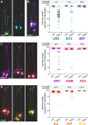
Figure 3.. Mutations in magi-1 preferentially affect URX and BAG dendrites.
Dendrite lengths in magi-1(zh66) animals were measured for (A, B) neurons that do not protrude into the external environment, but form specialized attachments (A) to the ILso glia: URX (green, flp-8pro) and BAG (blue, flp-17pro) or (B) to other glia: URY (dark purple, tol-1pro) (Doroquez et al., 2014; Ward et al., 1975); (C, D) neurons that protrude through glial pores and form tight and adherens junctions with glia: (C) amphid neurons AWC (light purple, odr-1pro), AWB (pink, str-1pro), ASG (red, ops-1pro) and (D) neurons in other sense organs, IL2 (dark orange, klp-6pro), CEP (light orange, dat-1pro), OLQ (yellow, ocr-4pro). Arrowheads, dendrite endings. Scale bars, 10 μm. Quantification of dendrite lengths is shown at right, expressed as a percentage of the distance from cell body to nose. Colored bars represent individual dendrites (n=50 per genotype); black bars represent population averages. Shaded region represents wild-type mean ± 5 SD for each neuron type and the percentage of dendrites in this range (“full length dendrites”) is indicated above the plots. Images from (A) and quantification of URX and BAG dendrite lengths are reproduced from Fig. 2.
Figure 4.
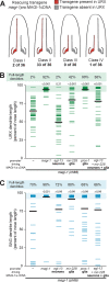
Figure 4.. MAGI-1 can act in glia to non-cell-autonomously promote URX dendrite extension.
(A) Schematic indicating results of a mosaic rescue experiment. Analysis was performed using magi-1(zh66) mutant animals bearing a stably integrated URX marker for assessing dendrite lengths (flp-8pro:GFP) and an unstable extrachromosomal array – stochastically lost during cell division – containing a rescuing MAGI-1 cDNA (magi-1pro:MAGI-1a) with a second URX marker for assessing the presence of the transgene in this neuron (flp-8pro:mCherry). To select for mosaicism, animals were selected in which only one of the two URX neurons was mCherry(+), indicating the presence of the rescuing cDNA, and the length of mCherry(+) (red) and mCherry(−) (gray) dendrites was assessed. The number of animals showing each of the four possible outcomes (Class I-IV) is indicated. (B-C) URX (B) and BAG (C) dendrite lengths, expressed as a percentage of the distance from the cell body to the nose, in magi-1(zh66) mutant animals bearing no transgene (−) or transgenes with a rescuing MAGI-1a cDNA under the control of the indicated promoters. Colored bars represent individual dendrites (n=50 per genotype); black bars represent population averages. Shaded region represents wild-type mean ± 5 SD and the percentage of dendrites in this range (“full length dendrites”) is indicated above the plots. magi-1(zh66) and +magi-1pro:MAGI-1 data are reproduced from Fig. 2. p-values, Kruskal-Wallis with Dunn’s post hoc test adusted for multiple comparisons.
Figure 5.
Figure 5.. MAGI-1 and SAX-7 directly bind each other, yet have partially independent functions in dendrite extension.
(A-B) Immunoblots from pull-down experiments. Glutathione S-transferase (GST) was fused to either the wild-type SAX-7 cytoplasmic tail (GST-SAX-7 WT) or the SAX-7 cytoplasmic tail lacking the PDZ-binding motif (GST-SAX-7∆PB). These purified components were used in pull-down experiments with lysates of HEK293T cells expressing full-length FLAG-tagged MAGI-1 (A) or purified bacterially-expressed HIS-tagged MAGI-1 (B). Following pull-down, MAGI-1 was detected by immunoblot (IB) for the FLAG or HIS epitopes and the GST-tagged SAX-7 cytoplasmic tail was detected by Ponceau S staining for total protein. Each pull-down was performed at least twice with similar results. (C) Schematic indicating the physical interaction between SAX-7 (blue) and MAGI-1 (green). Other hypothesized interactors are indicated in gray. (D) Images of magi-1(zh66) sax-7(qv30) L4 larvae expressing cell-specific reporters for URX (green, flp-8pro) and BAG (blue, flp-17pro) neurons. Anterior, up. The dashed line outlines the head. Green and blue arrowheads, URX and BAG dendrite endings, respectively. Scale bars, 10 μm. (E-F) URX (E) and BAG (F) dendrite lengths quantified as a percentage of the distance from the cell body to the nose in the indicated genotypes. Each colored bar represents a single dendrite (n=50 per genotype); black bars indicate population averages. The shaded region marks wild-type mean ± 5 SD, and the percentage of dendrites in this range (“full-length”) is indicated below the plots. Wild-type and single mutant data are reproduced from Fig. 1 and 2. p-values, Kruskal-Wallis with Dunn’s post hoc test adjusted for multiple comparisons.
Figure 6.
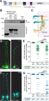
Figure 6.. The classical cadherin HMR-1 promotes URX and BAG dendrite extension.
(A,C) Images of URX (green, flp-8pro, A) and BAG (blue, flp-17pro, C) in wild-type (left) or sax-7(qv30) (right) animals with HMR-1 depleted from glia. Anterior, up. The dashed line outlines the head. Green and blue arrowheads, URX and BAG dendrite endings, respectively. Scale bars, 10 μm. Faint background fluorescent puncta are due to HMR-1-ZF1-GFP expression in non-glial cells. (B,D) URX (B) and BAG (D) dendrite lengths quantified as a percentage of the distance from the cell body to the nose in the indicated genotypes. “HMR-1 KD in glia” indicates knockdown of HMR-1 using the ZF1/ZIF-1 degron system for cell-specific degradation of HMR-1 using the pan-glial promoter mir-228pro. Each colored bar represents a single dendrite (n=50 per genotype); black bars indicate population averages. The shaded region marks wild-type mean ± 5 SD, and the percentage of dendrites in this range (“full-length”) is indicated below the plots. Wild-type and sax-7(qv30) data are reproduced from Fig. 1. Additional controls are shown in Supp. Fig. 2. (E) Immunoblot from pull-down experiments using purified recombinant components: the SAX-7 cytoplasmic tail fused to GST, HIS-tagged MAGI-1, and the HMP-2 PDZ-binding motif fused to MBP. Following pull-down, MAGI-1 and the HMP-2 motif were detected by immunoblot (IB) for the HIS or MBP epitopes respectively, and the GST-tagged SAX-7 cytoplasmic tail was detected by Ponceau S staining for total protein. Each pull-down was performed at least twice with similar results. (F) Schematic of MAGI-1 (green) bridging SAX-7 (blue) to the cadherin-catenin complex (gold) via HMP-2. p-values, Kruskal-Wallis with Dunn’s post hoc test adjusted for multiple comparisons.
Figure 7.
Figure 7.. The afadin homolog AFD-1 contributes to BAG dendrite extension.
(A) Diagram of AFD-1 indicating key protein domains and the allele used in this study. (B) Immunoblot showing pull-down experiments. GST was fused to either the wild-type SAX-7 cytoplasmic tail (GST-SAX-7 WT) or the SAX-7 cytoplasmic tail lacking the PDZ-binding motif (GST-SAX-7∆PB). These purified components were used in pull-down experiments with lysates of HEK293T cells expressing full-length FLAG-tagged AFD-1. Following pull-down, AFD-1 was detected by immunoblot (IB) for the FLAG epitope and the GST-tagged SAX-7 cytoplasmic tail was detected by Ponceau S staining for total protein. Each pull-down was performed at least twice with similar results. (C) Schematic indicating two non-mutually-exclusive mechanisms for bridging SAX-7 to MAGI-1: AFD-1 (orange) connects SAX-7 (teal) to MAGI-1 (lime green) or SAX-7 directly binds MAGI-1, which in turn interacts with the cadherin-catenin complex via binding to HMP-2 (yellow). (D,F) Representative images of URX (green, flp8pro, D) and BAG (blue, flp-17pro, F) afd-1(tm1250) (left) or afd-1(tm1250); magi-1(zh66) (right) mutant animals. Anterior, up. The dashed line outlines the head. Green arrowheads, URX dendrite endings. Blue arrowheads, BAG dendrite endings. Scale bars, 10 μm. (E,G) URX (E) and BAG (G) dendrite lengths quantified as a percentage of the distance from the cell body to the nose in the indicated genotypes. Each colored bar represents a single dendrite (n=50 per genotype); black bars indicate population averages. The shaded region marks wild-type mean ± 5SD, and the percentage of dendrites in this range (“full-length”) is indicated below the plots. magi-1(zh66) dendrite lengths are reproduced from Fig. 2. p-values, Kruskal-Wallis with Dunn’s post hoc test.

Similar articles

References

    1. Abedin M. and King N. (2010). Diverse evolutionary paths to cell adhesion. Trends Cell Biol 20, 734–742. - PMC - PubMed
    1. Adle-Biassette H., Saugier-Veber P., Fallet-Bianco C., Delezoide A.-L., Razavi F., Drouot N., Bazin A., Beaufrère A.-M., Bessières B., Blesson S., et al. (2013). Neuropathological review of 138 cases genetically tested for X-linked hydrocephalus: evidence for closely related clinical entities of unknown molecular bases. Acta Neuropathol 126, 427–442. - PubMed
    1. Armenti S. T., Lohmer L. L., Sherwood D. R. and Nance J. (2014). Repurposing an endogenous degradation system for rapid and targeted depletion of C. elegans proteins. Development 141, 4640–4647. - PMC - PubMed
    1. C. elegans Deletion Mutant Consortium (2012). large-scale screening for targeted knockouts in the Caenorhabditis elegans genome. G3 (Bethesda) 2, 1415–1425. - PMC - PubMed
    1. Cebul E. R., McLachlan I. G. and Heiman M. G. (2020). Dendrites with specialized glial attachments develop by retrograde extension using SAX-7 and GRDN-1. Development 147, dev180448. - PMC - PubMed

Publication types