[Preprint]. 2024 Jan 5:2024.01.04.574255.
doi: 10.1101/2024.01.04.574255.

Microvilli regulate the release modes of alpha-tectorin to organize the domain-specific matrix architecture of the tectorial membrane

Affiliations

Microvilli regulate the release modes of alpha-tectorin to organize the domain-specific matrix architecture of the tectorial membrane

Ava Niazi et al. bioRxiv. .

Abstract

The tectorial membrane (TM) is an apical extracellular matrix (ECM) in the cochlea essential for auditory transduction. The TM exhibits highly ordered domain-specific architecture. Alpha-tectorin/TECTA is a glycosylphosphatidylinositol (GPI)-anchored ECM protein essential for TM organization. Here, we identified that TECTA is released by distinct modes: proteolytic shedding by TMPRSS2 and GPI-anchor-dependent release from the microvillus tip. In the medial/limbal domain, proteolytically shed TECTA forms dense fibers. In the lateral/body domain produced by the supporting cells displaying dense microvilli, the proteolytic shedding restricts TECTA to the microvillus tip and compartmentalizes the collagen-binding site. The tip-localized TECTA, in turn, is released in a GPI-anchor-dependent manner to form collagen-crosslinking fibers, required for maintaining the spacing and parallel organization of collagen fibrils. Overall, we showed that distinct release modes of TECTA determine the domain-specific organization pattern, and the microvillus coordinates the release modes along its membrane to organize the higher-order ECM architecture.

Keywords: ECM; TECTA; TMPRSS2; alpha-tectorin; collagen; extracellular matrix; microvilli; tectorial membrane.


Figures
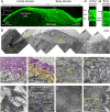
Figure 1.
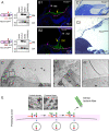
Figure 1.. The shapes of the apical cell surface and the associated matrix are distinct between the limbal and body domains of the developing tectorial membrane (TM).
A. PSA lectin staining of the TM at postnatal day (P) 2. The TM is composed of two domains along the radial axis: the limbal domain and the body domain. The limbal domain is produced by interdental cells and is composed of three layers: covernet (Cov), collagenous (Col), and border (Bor) layers. The developing body domain is produced by the greater epithelial ridge (GER) cells and is composed of two layers: covernet and central body (Cent Body) layers. PSA lectin staining shows distinct matrix architecture between the domains. M: medial; L: lateral. B. Transmission electron microscopy (TEM) of the TM at P2. b1: the most medial region of the limbal domain contains the extracellular vesicles (EVs; arrows) under the covernet layer (a pink area). b2: limbal domain. Dense non-collagenous fibers are present in all three layers of the limbal matrix (arrows). The apical surface of the interdental cells displays short and sparse microvilli. b3 and b3’: body domain. The apical surface of the GER cells displays dense and long microvilli. Radially oriented collagen fibrils are associated with the microvillus tip of the GER cells (pink curves). EVs are present (marked in light blue) near the microvillus-ECM border. I: the inner hair cell; O: the outer hair cell. C. TEM of the limbal matrix. Dense non-collagenous fibers (blue arrows) are not associated with individual collagen fibrils. Collagen fibrils (pink arrows) are not associated with crosslinking fibers and are less ordered. D. TEM of the body matrix. Collagen fibrils (pink arrows) are associated with crosslinking fibers (blue arrows), evenly spaced, and parallelly organized. E. Scanning electron microscopy (SEM) of the interdental cells (P2). The TM is removed genetically from the spiral limbus (Otoa knockout mice). Hexagonally arranged interdental cells contain medial microvilli on the apical junction. The non-junctional apical membrane displays sparse microvilli. F. SEM of the GER cells. The TM is removed by force during the tissue prep. The apical surface of the GER columnar cells displays densely packed microvilli. I: the inner hair cell.
Figure 2.
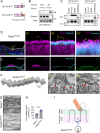
Figure 2.. TMPRSS2 sheds TECTA.
A. Diagram of TECTA and the ZP-domain of TECTA (TECTA-ZP). TECTA contains the NIDO domain, vWF domains, ZP domain, and C-terminal GPI-anchorage signal, which mediates collagen binding, ECM protein binding, polymerization, and GPI-anchorage, respectively. The ZP domain contains the consensus cleavage site (CCS) and the external hydrophobic patch (EHP) sequence close to the membrane-anchorage sequence. Cleavage of TMPRSS2 releases an ectodomain (red arrowheads), while cleavage by GPI-anchor lipases (blue arrows) releases non-proteolyzed protein. B. TECTA was expressed with an empty vector, GDE3 (a vertebrate GPI-anchor lipase) or TMPRSS2 in HEK293T cells, and the release of TECTA into the conditioned medium (CM) was monitored by western blot. The treatment of TECTA-expressing cells with phosphatidylinositol-phospholipase C (PI-PLC), a bacterial GPI-anchor lipase, or expression of GDE3 releases TECTA into the CM. TMPRSS2 releases a smaller fragment. Lys: lysate. C. Catalytic mutant of GDE3 (H230A) and TMPRSS2 (S441A) failed to release TECTA-ZP into the CM. PNGaseF was treated to both CM and Lys samples to show the size of the unglycosylated protein. D. Myc-TECTA-ZP and HA-TECTA-ZP were expressed with GDE3 or TMPRSS2. The released proteins in the CM were immunoprecipitated (IP) with Myc antibody and blotted with HA antibody. HA-TECTA-ZP released by TMPRSS2 was co-IPed by Myc antibody, while non-proteolyzed protein released by GDE3 was not. PNGaseF was treated to both CM and Lys samples to show the size of the unglycosylated protein. E. RNAscope in situ hybridization (ISH) of Tmrpss2 in the developing cochlea at P0. Tmprss2 (red) is expressed in both the spiral limbus (SL) and GER. Hoechst (blue). F. Immunohistochemistry (ISH) of TMPRSS2 in the developing cochlear at P2. The TMPRSS2 puncta were observed in the cytoplasm and apical area of the interdental cells (f1) and GER cells (f2). TMPRSS2 antibody staining requires antigen retrieval, which reduces phalloidin signal (see materials and methods). Annexin V (cyan) is enriched on the microvilli (von der Mark and Mollenhauer,1997) and used to label the apical surface of the GER cells. PSA (green); Hoechst (blue).
Figure 3.
Figure 3.. Proteolytic shedding of TECTA is required for the dense fiber formation in the limbal domain and the covernet layer.
A. R2061S (RS) mutation on CCS1 reduced the release of TECTA-ZP by TMPRSS2, while the release by PI-PLC or GDE3 remained intact. B. Airyscan images of PSA (green) and phalloidin (magenta) at P2. The limbal domain (magenta arrows) and the covernet layer (yellow arrows) are absent in the TM of TectaRS/RS mice. The body domain is associated with the GER cells but attached to the Reissner’s membrane (RM) in the absence of the covernet layer in TectaRS/RS mice. Hoechst (blue). C. Semi-thin section followed by toluidine blue staining of the adult cochlea at 4 weeks. In wildtype mice (C1), the mature TM is associated with the spiral limbus of the cochlea. In TectaRS/RS mice (C2), the TM is detached from the spiral limbus. D. TEM of the developing TM in TectaRS/RS mice at P2. The dense fibers in the limbal domain is absent in TectaRS/RS mice, resulting in the complete loss of the limbal domain (d2). The EV clusters in the most medium region remains intact (d1). E. Proposed model for formation of the dense tectorin fibers. Proteolytic shedding of TECTA is coupled with the elongation process of the ZP domain. Sequential process of the cleavage of the pre-assembled polymer and polymerization to the newly synthesized monomer will generate a long polymer species, which are stemming from the apical cell surface (3D printing model). The long polymer will form dense fiber in the limbal domain and covernet layer (red arrows).
Figure 4.
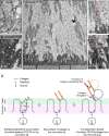
Figure 4.. Proteolytic cleavage of TECTA compartmentalizes the collagen-attachment site on the microvilli in the body domain.
A. TEM of the body domain of the wildtype at P2. Collagen fibrils are associated with the microvillus tip (marked in pink). B. TEM of TectaRS/RS homozygous mice at P2. Collagen fibrils are associated with the basal and lateral membrane of the microvilli (an arrow). The length of the microvilli is increased and variable. C. TEM of Tecta+/RS heterozygous mice at P2. Collagen fibrils are associated with the basal and lateral membrane of the microvilli (an arrow). Intermediate fibers (dead-end polymer) stemming from the microvillus membrane. D. Quantification of the microvillus length. Left graph: an average of the microvillus length per image was plotted. N=3 animals per group, 5 images per animal. Data was plotted as mean with SEM. Each symbol represents an individual image from three animals (magenta, green, and blue). Adjusted ∗∗∗∗p < 0.0001; **p < 0.01: *p < 0.05 was significant by Kruskal-Wallis test, Dunn’s multiple comparisons. E. Right graph: the length of individual microvilli was plotted. N=3 animals per group, n=215–259 microvilli per group. Data was plotted in a violin plot outlining the kernel probability density with the width of the shaded area representing the proportion of the data located there. The median and quartiles are also represented on the graph. F. Quantification of the microvillus density. The number of the microvilli along the horizontal apical surface was plotted. N=3 animals, 5 images per animal. Data was plotted as mean with SEM. Each symbol represents an individual image from animals (magenta, green, and blue). Adjusted p value was not significant by one-way ANOVA, Tukey’s multiple comparisons. G. TEM of the body matrix above the cellular border TectaRS/RS mice at P2. A large amount of EVs are accumulated in the lumen. H. TEM of the EVs in Tecta+/RS mice. EVs containing intermediated fibers are present near the microvilli. I. Airyscan IHC of TECTA showed that unlike wildtype TECTA, which is localized on the distal part of the microvilli (dashed curve), TECTARS is localized along the elongated microvilli. TECTARS is released from the cell and present within the matrix (an asterisk). J. Proposed model for the proteolytic cleavage of TECTA in the compartmentalization of the collagen-binding site. Proteolytic cleavage of TECTA removes excessive TECTA from the basal and lateral membrane of the microvillus. TECTA that reaches the microvillus tip may be protected from proteolytic shedding and recruit collagen fibrils. The EVs generated from the microvillus tip rapidly disintegrate. In TectaRS/RS mice, the proteolysis-resistant form of TECTA is accumulated on the lateral membrane, which results in the ectopic association of the collagen fibrils. Dysregulation of the matrix-association domain may lead to the instability of the microvillus architecture and over-stability of the EVs in the luminal space.
Figure 5.
Figure 5.. TECTA-ZP is enriched on the microvillus tip of W4 cells in the presence of TMPRSS2.
A. LS174T-W4 cells (colon cancer cells) were transfected with TECTA-ZP with and without TMPRSS2 and the localization of the surface TECTA-ZP (sTECTA-ZP, magenta) and EPS8 (green), a microvillus tip marker, were measured. Doxycycline (DOX) treatment (1 μg/ml, 16 hours), which activates the polarity kinase LKB1, induces the accumulation of EPS8. sTECTA-ZP is co-localized with EPS8 in the presence of TMPRSS2 expression. TMPRSS2 expression did not induce the colocalization of the cleavage-resistant form (TECTA-ZPRS) with EPS8. Hoechst (blue). K. Quantification of the percentage of sTECTA-ZP colocalized with EPS8. The colocalization was measured as the percentage of the integrated intensity of sTECTA-ZP (number of the pixels*average pixel intensity) in the overlapped ROI over the total ROI. Data was plotted as mean with SEM. Each symbol represents an individual image from three pooled experiments (N=3, n=5 images per experimental condition). Adjusted ∗∗∗p < 0.001, or ns, not significant by one-way ANOVA, Tukey’s multiple comparisons.
Figure 6.
Figure 6.. GPI-anchorage of TECTA is required for the release of TECTA from the microvillus tip.
A. Diagram of the TECTA-ZP wildtype and swapping mutant with a transmembrane domain of ZP3. B. Surface biotinylation of TECTA-wildtype (WT) and TECTA-ZP3 swapping mutant expressed in HEK293T cells. TECTA-ZP was expressed on the cell surface membrane and not released by PI-PLC treatment, while TECTA-WT was removed from the cell surface and released into the CM. C. TMPRSS2 expression released TECTA-ZPZP3 from HEK293T cells to the CM. GPI-lipases (PI-PLC treatment or GDE3 expression) did not induce the release of TECTA-ZPZP3. D. Airyscan image of TECTA (magenta), phalloidin (cyan), PSA lectin (green), and Hoechst (blue) of the TM in TectaZP3/ZP3 mice at P2. of TECTA-ZPZP3 is localized on the microvillus tip (arrows). The release of TECTA-ZPZP3 was dramatically reduced (an asterisk). E. Airyscan image of collagen II (Col2: magenta), phalloidin (cyan), PSA lectin (green), and Hoechst (blue) of the TM in TectaZP3/ZP3 mice at P2. Collagen II is assembled in the TM and associated with the apical surface in TectaZP3/ZP3 mice. F. TEM of the TM in TectaZP3/ZP3 mice at P2. Collagen fibrils are associated with the microvillus tip of TectaZP3/ZP3 mice (arrows). Collagen fibrils displaying a loop-like association at both ends were observed (an asterisk). G. TEM of the collagen fibrils of the body matrix. In wildtype mice, collagen fibrils are interconnected with collagen-crosslinking fibers and evenly spaced. Collagen fibrils of the TectaZP3/ZP3 mice lack crosslinking fibers and are condensed. H. The number of crossing of collagen fibrils along the perpendicular line is increased in TectaZP3/ZP3 mice. Data are plotted as mean with SEM. The number of the collagen fibrils across a 1 μm line perpendicular to the fibers was manually counted and plotted. N=3 animals per group, 5 images per animal. Each symbol represents an individual image from three animals (magenta, green and blue). Adjusted ∗∗∗∗p < 0.0001, by unpaired Student’s t-test. I. Diagram of the TECTA localization and collagen arrangement in TectaZP3/ZP3 mice. TECTA-ZPZP3 is normally processed by proteases on the basal and lateral microvillus membrane and enriched on the microvillus tip. The tip-localized TECTA-ZPZP3 recruits collagen fibrils. Without GPI-anchorage, TECTA is not released into the matrix, which results in the absence of collagen-crosslinking fibers. Collagen fibrils lacking crosslinking fibers are condensed and disorganized.
Figure 7.
Figure 7.. EVs are produced from the microvilli of the GER cells.
A. TEM of the microvilli in wildtype mice at P2. Budding of the vesicles from the microvillus tip was observed. B. TEM of the lumen in TectaRS/RS mice at P2. A large amount of EVs was accumulated in the lumen. C. TEM of the TectaRS/ZP3 mice at P2. EV-like circular clusters were observed along the collagen fibrils that are connected to the microvillus tip. D. Proposed model for the role of microvilli in collagen organization. Newly synthesized TECTA is delivered to the base of the microvilli and migrates to the microvillus tip along the membrane. Excessive collagen is removed from the basal and lateral membrane by proteolytic shedding. The shed TECTA ectodomain may form a dimer or a small oligomer with surface-tethered TECTA and migrate to the microvillus tip (1) or travel in the extracellular space and be captured on the microvillus tip (2). TECTA that reaches the microvillus tip (safe zone) is no longer processed and is not organized into a long polymer (dense tectorin fibers). The tip-localized TECTA recruits collagen fibrils. TECTA/collagen complex is released from the tip via EVs. The released EVs rapidly disintegrate, and dispersed TECTA forms the collagen-crosslinking fibers. The budding of EVs may expose the end of the collagen fibrils so a new collagen molecule (outlined in pink) is incorporated into the growing end.

Similar articles

References

    1. Frantz C., Stewart K.M., and Weaver V.M. (2010). The extracellular matrix at a glance. J Cell Sci 123, 4195–4200. 10.1242/jcs.023820. - DOI - PMC - PubMed
    1. Peixoto C.A., and De Souza W. (1995). Freeze-fracture and deep-etched view of the cuticle of Caenorhabditis elegans. Tissue Cell 27, 561–568. - PubMed
    1. Richardson S.H., Starborg T., Lu Y., Humphries S.M., Meadows R.S., and Kadler K.E. (2007). Tendon development requires regulation of cell condensation and cell shape via cadherin-11-mediated cell-cell junctions. Mol Cell Biol 27, 6218–6228. 10.1128/MCB.00261-07. - DOI - PMC - PubMed
    1. Chen S., Mienaltowski M.J., and Birk D.E. (2015). Regulation of corneal stroma extracellular matrix assembly. Exp Eye Res 133, 69–80. 10.1016/j.exer.2014.08.001. - DOI - PMC - PubMed
    1. Mouw J.K., Ou G., and Weaver V.M. (2014). Extracellular matrix assembly: a multiscale deconstruction. Nat Rev Mol Cell Biol 15, 771–785. 10.1038/nrm3902. - DOI - PMC - PubMed

Publication types