. 2024 Jan 23;81(1):55.
doi: 10.1007/s00018-023-05088-w.

Ribosomal protein L24 mediates mammalian microRNA processing in an evolutionarily conserved manner

Affiliations

Ribosomal protein L24 mediates mammalian microRNA processing in an evolutionarily conserved manner

Yonat Tzur et al. Cell Mol Life Sci. .

Abstract

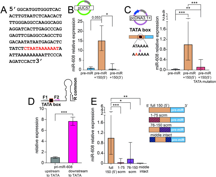
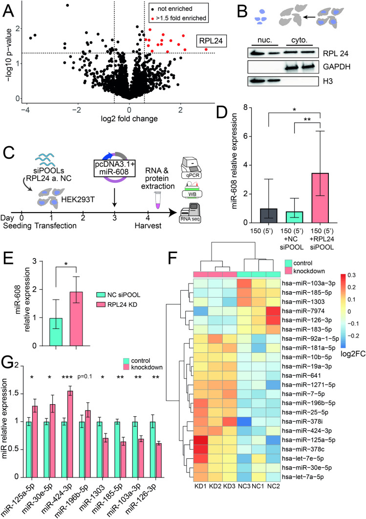
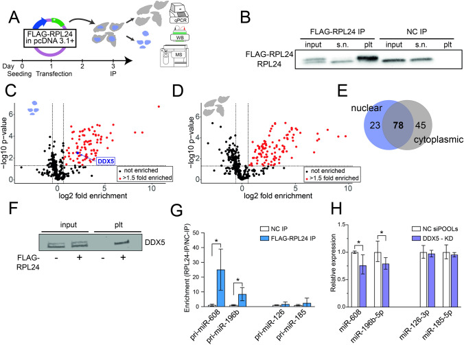
To investigate the mechanism(s) underlying the expression of primate-specific microRNAs (miRs), we sought DNA regulatory elements and proteins mediating expression of the primate-specific hsa-miR-608 (miR-608), which is located in the SEMA4G gene and facilitates the cholinergic blockade of inflammation by targeting acetylcholinesterase mRNA. 'Humanized' mice carrying pre-miR-608 flanked by 250 bases of endogenous sequences inserted into the murine Sema4g gene successfully expressed miR-608. Moreover, by flanking miR-608 by shortened fragments of its human genome region we identified an active independent promoter within the 150 nucleotides 5' to pre-miR-608, which elevated mature miR-608 levels by 100-fold in transfected mouse- and human-originated cells. This highlighted a regulatory role of the 5' flank as enabling miR-608 expression. Moreover, pull-down of the 150-base 5' sequence revealed its interaction with ribosomal protein L24 (RPL24), implicating an additional mechanism controlling miR-608 levels. Furthermore, RPL24 knockdown altered the expression of multiple miRs, and RPL24 immunoprecipitation indicated that up- or down-regulation of the mature miRs depended on whether their precursors bind RPL24 directly. Finally, further tests showed that RPL24 interacts directly with DDX5, a component of the large microprocessor complex, to inhibit miR processing. Our findings reveal that RPL24, which has previously been shown to play a role in miR processing in Arabidopsis thaliana, has a similar evolutionarily conserved function in miR biogenesis in mammals. We thus characterize a novel extra-ribosomal role of RPL24 in primate miR regulation.

Keywords: DDX5; Hsa-miR-608; Pre-miRs; Pri-miRs; RPL24; miR processing.


Conflict of interest statement

The authors declare no competing interests.

Figures

Fig. 1
Fig. 1
Cis-sequences regulate miR-608 expression in engineered KI mice and cultured cells. A Structure of the mouse construct: miR-608 transgenic ‘humanized’ mice were established by inserting hsa-pre-mir-608 into the third intron of the mouse Sema4g gene, with 250 flanking bases at each side. B miR-608 levels in the brain and peripheral tissues of female and male miR-608 KI mice, determined by RT-qPCR. All miR-608 levels were normalized to those of female hypothalamus (that showed the lowest expression); two-way ANOVA with Dunnet’s multiple comparisons correction, ± SD, n = 5 per group. C Experimental design: HEK293T and CT26.WT cells were seeded and 24 h later transfected with pcDNA3.1 + plasmid containing miR-608 inserted into the second intron of HBB and flanked by native sequences of varying lengths. Cells were harvested 48 h later, RNA extracted, and miR-608 levels quantified. D Symmetric bidirectional 250 base extension of the miR-608 stem-loop altered its levels in HEK293T cells; bar graph, ± SD, p = 0.0163, unpaired t-test. E Constructs of pre-miR-608 flanked by symmetric sequences ranging from 75 to 250 bases. F Levels of miR-608 expressed from these constructs in HEK293T cells. The 150 bases both upstream and downstream are critical for miR-608 expression in HEK293T cells. G The 150-base symmetrical flanks are also critical for expression in CT26.WT cells. H Constructs of pre-miR-608 flanked by asymmetric sequences ranging from 75 to 150 bases. I Levels of miR-608 expressed from these constructs in HEK293T cells were highest under control of the upstream 150 base sequence. J A similar effect is seen in CT26.WT cells transfected with these constructs. All experiments were performed in duplicate or triplicate and miR-608 levels were measured using Taqman RT-qPCR with RNU6B and snoRNA135 as normalizing genes. Results are shown relative to levels of pre-miR-608 with no flanking sequences. In all panels *p < 0.05, **p < 0.01, ***p < 0.001. In panels F, G, I, bar-graph ± SD, one-way ANOVA with Tukey’s multiple comparisons test
Fig. 2
Fig. 2
A TATA box enabling miR-608 expression is located in the 5′ 150 bases. A The 150 bases 5′ upstream to pre-miR-608 contain a TATA box at position 111 (red). B miR-608 levels in HEK293T cells transfected with pUC57, quantified by Taqman RT-qPCR, show that the TATA box drives expression from the promoter-less bacterial vector pUC57. C. miR-608 levels in HEK293T cells transfected with the mammalian vector pcDNA3.1 + containing the TATA box T-to-A point mutation are decreased as compared to non-mutated TATA box. D Higher levels of pri-miR-608 transcripts (expressed from pcDNA3.1 +) and measured by RT-qPCR using forward primers positioned upstream (F1) vs downstream (F2) to the predicted TATA box; the reverse primer (R) is common. E Scrambling parts of the 5′ 150 bases upstream to the pre-miR-608 sequence decreased the levels of mature miR-608, as determined by Taqman RT-qPCR quantification of miR-608 relative to the intact 150 base sequence and normalized to RNU6B. Hatching represents the scrambled area. Experiments were performed in duplicate or triplicate and in all panels *p < 0.05, **p < 0.01, ***p < 0.001. In panel D unpaired t-test, in all other panels one-way ANOVA with Tukey’s correction for multiple comparisons, bar-graph ± SD
Fig. 3
Fig. 3
RPL24 KD alters the levels of diverse miRs in addition to miR-608. A Pull-down analysis: mass spectrometry identified 15 proteins bound to the 5′ 150 base-upstream sequence with p < 0.05 and enrichment > 1.5-fold (marked in red), the most enriched being RPL24 (p = 0.039, fold enrichment = 8). B Immunoblot of subcellular fractions identified RPL24 in both the nuclear and the cytoplasmic fractions. Fraction purity was validated with GAPDH, a cytoplasmic marker, and H3, a nuclear marker; each of which was localized solely to its expected compartment. C Experimental design: HEK293T cells were seeded, transfected with 50 nM of siPOOLs targeting RPL24 or a non-targeting negative control pool (NC) 24 h later, then transfected with pcDNA3.1 + containing miR-608 48 h later. RNA and proteins were extracted 24 h post-transfection. D RPL24 depletion in HEK293T cells elevates miR-608 levels. E RPL24 depletion in Caco2 cells shows the same effect. Quantification of miR-608 by Taqman RT-qPCR, with results normalized to RNU6B and relative to NC siPOOLs; for D, one-way ANOVA with Tukey’s correction for multiple comparisons ± SD. For E, unpaired t-test ± SD. F Heatmap showing the 22 DE miRs increased or decreased after RPL24 KD, analyzed by DESeq2, adjusted p-value < 0.05, Benjamini–Hochberg correction [27]. See Supp. Table 3 for full information on the DE miRs. G RT-qPCR validation confirming the RNA-seq results with data shown relative to the NC and normalized to SNORD47 and SNORD48; unpaired t-test for each miR ± SD, in panels D, E, G, *p < 0.05, **p < 0.01, ***p < 0.001
Fig. 4
Fig. 4
RPL24 binds pri-miRs and inhibits their processing through direct interaction with DDX5. A Experimental design: HEK293T cells were seeded and 24 h later transfected with pcDNA3.1 + vector containing FLAG-RPL24 or no insert as control. Cells were harvested 48 h after transfection, fractionated, and nuclear and cytoplasmic fractions immunoprecipitated separately with an antibody against the FLAG-tag, with subsequent MS analysis. B Immunoblot of nuclear fraction from cells transfected with a pcDNA3.1 + vector containing FLAG-RPL24 insert or empty pcDNA3.1 + vector as control, showing pull-down of the tagged-RPL24. 2.5% of input lysate, 2.5% of supernatant, and 20% of pellet were loaded per lane. Of the double band observed in the input lane, the upper and lower bands are the FLAG-RPL24 (~ 24kDa) and the endogenous RPL24 (~ 23kDa). C, D Volcano plot presenting the proteins enriched in FLAG-RPL24 IP compared to control, in the nuclear (C) and cytoplasmic (D) fractions. Red symbols denote proteins with p < 0.05 and enrichment > 1.5-fold and black symbols denote non-significant proteins. E Venn diagram showing proteins bound to RPL24 in cytoplasmic and nuclear fractions, with common and specific proteins. F Immunoblot showing the presence of DDX5 in RPL24 IP from the nuclear fraction of HEK293T cells transfected with pcDNA3.1 + vector containing ( +) vs lacking (−) a FLAG-RPL24 insert, confirming RPL24-DDX5 interaction. 2.5% of input lysate and 90% of pellet were loaded per lane. G RT-qPCR quantification of pri-miRs in nuclear pellet samples of FLAG-RPL24 IP. Pri-miR-608 and pri-miR-196b are enriched, but pri-miR-126 and pri-miR-185 are not. Each pellet sample was normalized to its corresponding input sample and fold enrichment is determined as FLAG-RPL24 IP/NC IP. H RT-qPCR quantification of miRs following DDX5 KD. miR-608 and miR-196b-5p are downregulated, but miR-126-3p and miR-185-5p levels are unchanged. For Panels G and H, experiments were performed in triplicate. Unpaired t-test, bar-graph ± SD, *p < 0.05

Similar articles

Cited by

References

    1. Bartel DP. Metazoan MicroRNAs. Cell. 2018;173(1):20–51. doi: 10.1016/j.cell.2018.03.006. - DOI - PMC - PubMed
    1. Barbash S, Shifman S, Soreq H. Global coevolution of human microRNAs and their target genes. Mol Biol Evol. 2014;31(5):1237–1247. doi: 10.1093/molbev/msu090. - DOI - PubMed
    1. Fukuda T, et al. DEAD-box RNA helicase subunits of the Drosha complex are required for processing of rRNA and a subset of microRNAs. Nat Cell Biol. 2007;9(5):604–611. doi: 10.1038/ncb1577. - DOI - PubMed
    1. Kim YK, Kim B, Kim VN. Re-evaluation of the roles of DROSHA, Export in 5, and DICER in microRNA biogenesis. Proc Natl Acad Sci U S A. 2016;113(13):E1881–E1889. doi: 10.1073/pnas.1602532113. - DOI - PMC - PubMed
    1. Ha M, Kim VN. Regulation of microRNA biogenesis. Nat Rev Mol Cell Biol. 2014;15(8):509–524. doi: 10.1038/nrm3838. - DOI - PubMed