

Advancement in pleura effusion diagnosis: a systematic review and meta-analysis of point-of-care ultrasound versus radiographic thoracic imaging
- PMID: 38261109
- PMCID: PMC10805747
- DOI: 10.1186/s13089-023-00356-z
Advancement in pleura effusion diagnosis: a systematic review and meta-analysis of point-of-care ultrasound versus radiographic thoracic imaging
Abstract
Background: Pleural effusion is a fluid buildup in the pleural space that mostly result from congestive heart failure, bacterial pneumonia, malignancy, and pulmonary embolism. The diagnosis of this condition can be challenging as it presents symptoms that may overlap with other conditions; therefore, imaging diagnostic tools such as chest x-ray/radiograph (CXR), point-of-care ultrasound (POCUS), and computed tomography (CT) have been employed to make an accurate diagnosis. Although POCUS has high diagnostic accuracy, it is yet to be considered a first-line diagnostic tool as most physicians use radiography. Therefore, the current meta-analysis was designed to compare POCUS to chest radiography.
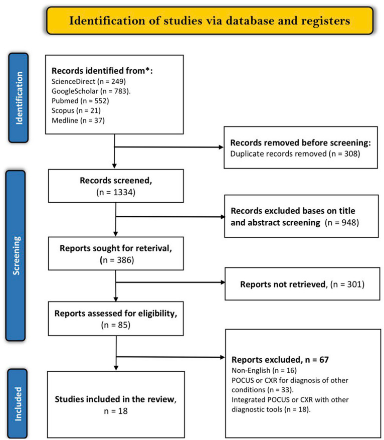
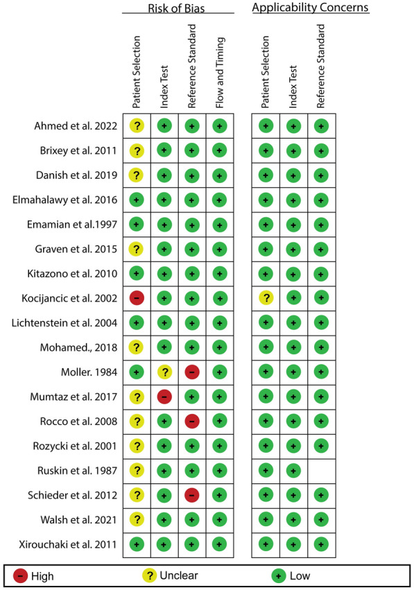
Methods: n extended search for studies related to our topic was done on five electronic databases, including PubMed, Medline, Embase, Scopus, and Google Scholar. A quality assessment using the Quality Assessment of Diagnostic Accuracy Studies tool (QUADAS-2) was performed on all eligible articles obtained from the databases. Moreover, the diagnostic accuracy of POCUS and CXR was performed using STATA 16 software.
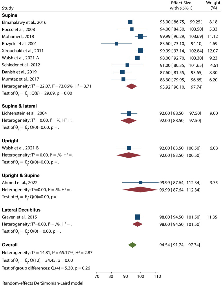
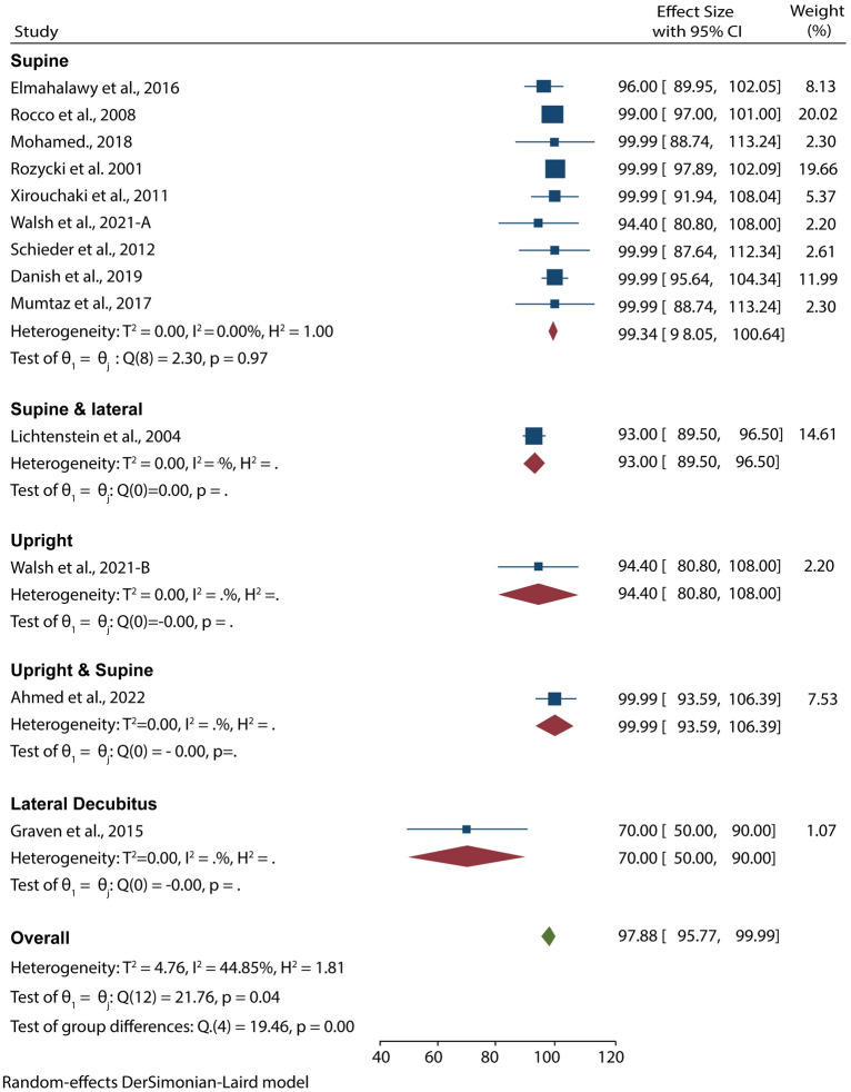
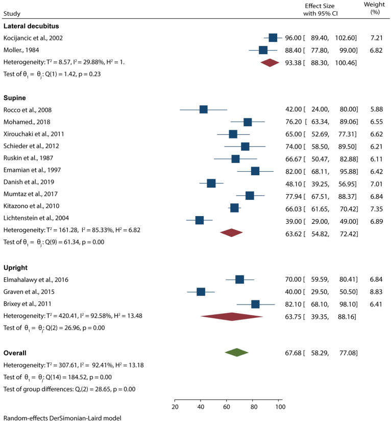
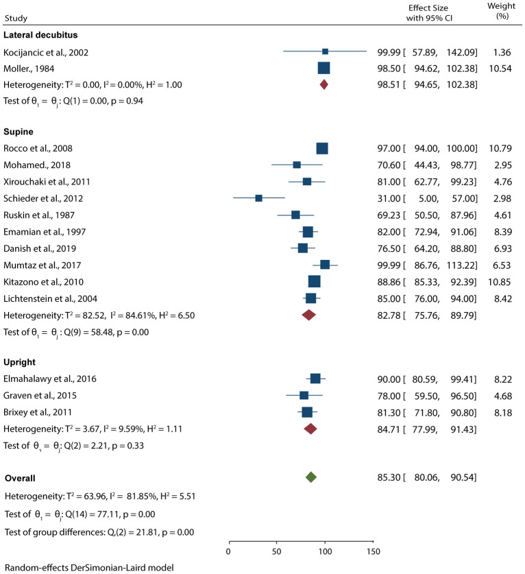
Results: Our search yielded 1642 articles, of which only 18 were eligible for inclusion and analysis. The pooled analysis showed that POCUS had a higher diagnostic accuracy compared to CXR (94.54% (95% CI 91.74-97.34) vs. 67.68% (95% CI 58.29-77.08) and 97.88% (95% CI 95.77-99.99) vs. 85.30% (95% CI 80.06-90.54) sensitivity and specificity, respectively). A subgroup analysis based on the position of patients during examinations showed that POCUS carried out in supine and upright positions had higher specificity than other POCUS positions (99%). In comparison, lateral decubitus CXR had higher sensitivity (96%) and specificity (99%) than the other CXR positions. Further subgroup analyses demonstrated that CXR had higher specificity in studies that included more than 100 patients (92.74% (95% CI 85.41-100). Moreover, CXR tends to have a higher diagnostic accuracy when other CXR positions are used as reference tests (93.38% (95% CI 86.30-100) and 98.51% (95% CI 94.65-100) sensitivity and specificity, respectively).
Conclusion: POCUS as an imaging modality has higher diagnostic accuracy than CXR in detecting pleural effusion. Moreover, the accuracy is still high even when performed by physicians with less POCUS training. Therefore, we suggest it is considered a first-line imaging tool for diagnosing pleural effusion at the patients' bedside.
Keywords: Chest X-ray; Diagnostic imaging; Emergency medicine; Meta-analysis; Pleural effusion; Point-of-care systems; Sensitivity; Specificity; Systematic review; Ultrasound.
© 2024. The Author(s).
Conflict of interest statement
No conflict of interest by the authors to declare.
Figures
Similar articles
-
Chest ultrasonography versus supine chest radiography for diagnosis of pneumothorax in trauma patients in the emergency department.Cochrane Database Syst Rev. 2020 Jul 23;7(7):CD013031. doi: 10.1002/14651858.CD013031.pub2. Cochrane Database Syst Rev. 2020. PMID: 32702777 Free PMC article.
-
Deep venous thrombosis (DVT) diagnostics: gleaning insights from point-of-care ultrasound (PoCUS) techniques in emergencies: a systematic review and meta-analysis.Ultrasound J. 2024 Jul 30;16(1):37. doi: 10.1186/s13089-024-00378-1. Ultrasound J. 2024. PMID: 39080184 Free PMC article. Review.
-
Diagnostic Accuracy of Point-of-Care Lung Ultrasonography and Chest Radiography in Adults With Symptoms Suggestive of Acute Decompensated Heart Failure: A Systematic Review and Meta-analysis.JAMA Netw Open. 2019 Mar 1;2(3):e190703. doi: 10.1001/jamanetworkopen.2019.0703. JAMA Netw Open. 2019. PMID: 30874784 Free PMC article.
-
Integrating point-of-care ultrasound in the ED evaluation of patients presenting with chest pain and shortness of breath.Am J Emerg Med. 2019 Feb;37(2):298-303. doi: 10.1016/j.ajem.2018.10.059. Epub 2018 Oct 30. Am J Emerg Med. 2019. PMID: 30413369
-
Diagnostic value of pleural ultrasound to refine endotracheal tube placement in pediatric intensive care unit.Arch Pediatr. 2021 Nov;28(8):712-717. doi: 10.1016/j.arcped.2021.09.006. Epub 2021 Oct 6. Arch Pediatr. 2021. PMID: 34625381
Cited by
-
Diagnostic and Therapeutic Approach in Pediatric Pulmonary Abscess: Two Cases and Literature Review.J Clin Med. 2024 Dec 20;13(24):7790. doi: 10.3390/jcm13247790. J Clin Med. 2024. PMID: 39768711 Free PMC article. Review.
-
A Practical Approach to Pleural Infection.Pulm Ther. 2025 Sep;11(3):535-551. doi: 10.1007/s41030-025-00308-z. Epub 2025 Jul 25. Pulm Ther. 2025. PMID: 40715732
-
Advanced imaging techniques and artificial intelligence in pleural diseases: a narrative review.Eur Respir Rev. 2025 Apr 2;34(176):240263. doi: 10.1183/16000617.0263-2024. Print 2025 Apr. Eur Respir Rev. 2025. PMID: 40174960 Free PMC article. Review.
-
Neonatal point-of-care ultrasound-guidelines for training, credentialing and quality assurance.J Perinatol. 2025 Aug 1. doi: 10.1038/s41372-025-02367-1. Online ahead of print. J Perinatol. 2025. PMID: 40751094 Review.
-
Pleural Infection: Diagnosis, Management, and Future Directions.J Clin Med. 2025 Mar 2;14(5):1685. doi: 10.3390/jcm14051685. J Clin Med. 2025. PMID: 40095674 Free PMC article. Review.
References
-
- Boka K (2022) Pleural Effusion: Background, Anatomy, Etiology. Published Online First. https://emedicine.medscape.com/article/299959-overview#a9
Publication types
LinkOut - more resources
Full Text Sources