Review
. 2024 Jan 23;166(1):28.
doi: 10.1007/s00701-024-05910-9.

Grading meningioma resections: the Simpson classification and beyond

Affiliations
Review

Grading meningioma resections: the Simpson classification and beyond

Matthias Simon et al. Acta Neurochir (Wien). .

Abstract

Technological (and also methodological) advances in neurosurgery and neuroimaging have prompted a reappraisal of Simpson's grading of the extent of meningioma resections. To the authors, the published evidence supports the tenets of this classification. Meningioma is an often surgically curable dura-based disease. An extent of meningioma resection classification needs to account for a clinically meaningful variation of the risk of recurrence depending on the aggressiveness of the management of the (dural) tumor origin.Nevertheless, the 1957 Simpson classification undoubtedly suffers from many limitations. Important issues include substantial problems with the applicability of the grading paradigm in different locations. Most notably, tumor location and growth pattern often determine the eventual extent of resection, i.e., the Simpson grading does not reflect what is surgically achievable. Another very significant problem is the inherent subjectivity of relying on individual intraoperative assessments. Neuroimaging advances such as the use of somatostatin receptor PET scanning may help to overcome this central problem. Tumor malignancy and biology in general certainly influence the role of the extent of resection but may not need to be incorporated in an actual extent of resection grading scheme as long as one does not aim at developing a prognostic score. Finally, all attempts at grading meningioma resections use tumor recurrence as the endpoint. However, especially in view of radiosurgery/radiotherapy options, the clinical significance of recurrent tumor growth varies greatly between cases.In summary, while the extent of resection certainly matters in meningioma surgery, grading resections remains controversial. Given the everyday clinical relevance of this issue, a multicenter prospective register or study effort is probably warranted (including a prominent focus on advanced neuroimaging).

Keywords: Extent of resection; Meningioma; Meningioma surgery; Simpson grading.


Figures

Fig. 1
Fig. 1
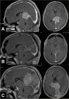
A September 2021: a large right occipital meningioma diagnosed in a 69-year-old woman (atypical meningioma WHO/CNS grade II). B December 2021: MR scans obtained 2 weeks after removal of an infected bone flap and epidural abscess. A small contrast-enhancing structure was interpreted as reactive tissue but in retrospect likely was residual tumor (yellow arrow). C October 2022: large recurrence likely originating from the small meningioma remnant
Fig. 2
Fig. 2
A and B, C Two cases with convexity meningiomas. Resecting the dural tumor origin of a convexity tumor is particularly easy. In the second case (B, C), there is a somewhat prominent “dural tail” (yellow arrows). At surgery, there was prominent dural hypervascularity but also a thin dural tumor layer surrounding the actual tumor mass. The latter observation together with the finding of microscopic dura infiltration [6, 7, 23] may argue for removal of “dural tails” in order to achieve a complete tumor resection. Of note (and as in our case), not all dural contrast enhancement depicted by MR scanning corresponds to actual tumor invasion (and not all invasion is detected by MRI). Incidence and extent of meningioma infiltration probably vary with the radiological characteristics of the “dural tail” [1, 47, 55]. D, E A small 15 mm meningioma originating from the lower border of the frontal falx. Simpson grade I resections of meningiomas of the falx are usually possible at little if any additional risk to the patient as long as there is some distance to the superior sagittal sinus. Infiltration of the superior sagittal sinus in falcine and convexity “parasagittal” meningiomas may preclude Simpson grade I, II and even III resections. It is however usually safely possible to resect the edge of the sinus, and one can follow the tumor into the sinus for removal of smaller intrasinusoidal fragments [18, 34]. F Intraventricular meningiomas have no true “dural” origin but rather originate from arachnoid cap cell populations in the choroidal plexus. The need to obtain hemostasis necessitates coagulation and removal of parts of the plexus, i.e., most of the times the surgery quite automatically results in a Simpson grade I resection
Fig. 3
Fig. 3
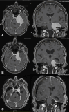
A Atypical falcotentorial meningioma WHO/CNS grade II operated following a first generalized seizure in a 20-year-old male. B Postoperative MR imaging confirmed the intraoperative impression of a Simpson grade III resection. C The patient did not report back to our center until 53 months later. At this time, there was a very large recurrence likely originating from the tumor infiltrated dura left behind during the initial surgery. However, resecting the dural tumor origin, i.e., the sinuses involved in this case during the first operation would not have been safe
Fig. 4
Fig. 4
A A large olfactory groove meningioma with B, C infiltration of the bony skull base (yellow arrow). Resection of the tumor infiltrated dura and bone (Simpson grade I) may be a bit cumbersome and result in the need of reconstructing the skull base using, e.g., a periosteal flap, but does not add much risk to the operation
Fig. 5
Fig. 5
In most cases with skull base menigiomas, it may be technically very challenging to achieve even a Simpson grade III resection. A Depending on the specifics of cavernous sinus involvement the dural tumor origin of medial sphenoid wing and anterior clinoidal meningiomas can sometimes be resected. We do not routinely enter the cavernous sinus and at most remove its outer dural layer. B In cases with tuberculum sellae/planum sphenoidale meningiomas, tumor infiltration of the diaphragma sellae and the optic canals may limit the aggressiveness of the resection. C We usually do not resect the tentorial edge or enter the cavernous sinus (yellow arrow). D In clival and E, F petroclival meningiomas, resections are limited by the necessary manipulation of the brainstem, cranial nerves, and their vasculature. The cases shown had some coagulation of the dural tumor origins (i.e., Simpson grade II/III) resections
Fig. 6
Fig. 6
In some cases, it may be best to aim for an incomplete (staged) resection combined with later radiosurgery. A Large spheno-petro-clival meningioma diagnosed in a 46-year-old female presenting with a seizure. B The supratentorial part of the tumor was removed through a transsylvian-subtemporal route. C The patient had a second surgery 3 months later for resection of the infratentorial tumor using a lateral suboccipital route. Similar to others, we often prefer staged surgery and standard rather than true one-stage skull base (e.g., transpetrosal) approaches [15, 50]
Fig. 7
Fig. 7
Spinal meningiomas most often originate from the antero-lateral dura. The authors remove the inner dural layer (“Simpson grade 1.5”) in such cases and try to avoid dural resections and reconstructions which require spinal cord traction and carry a significant risk of incurring CSF fistulas

References

    1. Ahmadi J, Hinton DR, Segall HD, Couldwell WT (1994) Surgical implications of magnetic resonance-enhanced dura. Neurosurgery 35:370–377. 10.1227/00006123-199409000-00003 - PubMed
    1. Baumgarten P, Gessler F, Schittenhelm J, Skardelly M, Tews DS, Senft C, Dunst M, Imoehl L, Plate KH, Wagner M, Steinbach JP, Seifert V, Harter PN (2016) Brain invasion in otherwise benign meningiomas does not predict tumor recurrence. Acta Neuropathol 132:479–481 - PubMed
    1. Behling F, Fodi C, Hoffmann E, Renovanz M, Skardelly M, Tabatabai G, Schittenhelm J, Honegger J, Tatagiba M (2021) The role of Simpson grading in meningiomas after integration of the updated WHO classification and adjuvant radiotherapy. Neurosurg Rev 44:2329–2336. 10.1007/s10143-020-01428-7 - PMC - PubMed
    1. Behling F, Bruneau M, Honegger J, Berhouma M, Jouanneau E, Cavallo L, Cornelius JF, Messerer M, Daniel RT, Froelich S, Mazzatenta D, Meling T, Paraskevopoulos D, Roche P-H, Schroeder HWS, Zazpe I, Voormolen E, Visocchi M, Kasper E, Schittenhelm J, Tatagiba M (2023) Differences in intraoperative sampling during meningioma surgery regarding CNS invasion - results of a survey on behalf of the EANS skull base section. Brain Spine 3:101740. 10.1016/j.bas.2023.101740 - PMC - PubMed
    1. Bernat AL, Priola SM, Elsawy A, Farrash F, Pasarikovski CR, Almeida JP, Lenck S, De Almeida J, Vescan A, Monteiro E, Zadeh GM, Gentili F (2018) Recurrence of anterior skull base meningiomas after endoscopic endonasal resection: 10 years’ experience in a series of 52 endoscopic and transcranial cases. World Neurosurg 120:e107-e113. 10.1016/j.wneu.2018.07.210 - PubMed

Publication types

LinkOut - more resources