. 2024 Jan 2;65(1):40.
doi: 10.1167/iovs.65.1.40.

Effector T Cells Promote Fibrosis in Corneal Transplantation Failure

Affiliations

Effector T Cells Promote Fibrosis in Corneal Transplantation Failure

Shudan Wang et al. Invest Ophthalmol Vis Sci. .

Abstract

Purpose: To evaluate whether fibrosis contributes to corneal transplant failure and to determine whether effector CD4+ T cells, the key immune cells in corneal transplant rejection, play a direct role in fibrosis formation.

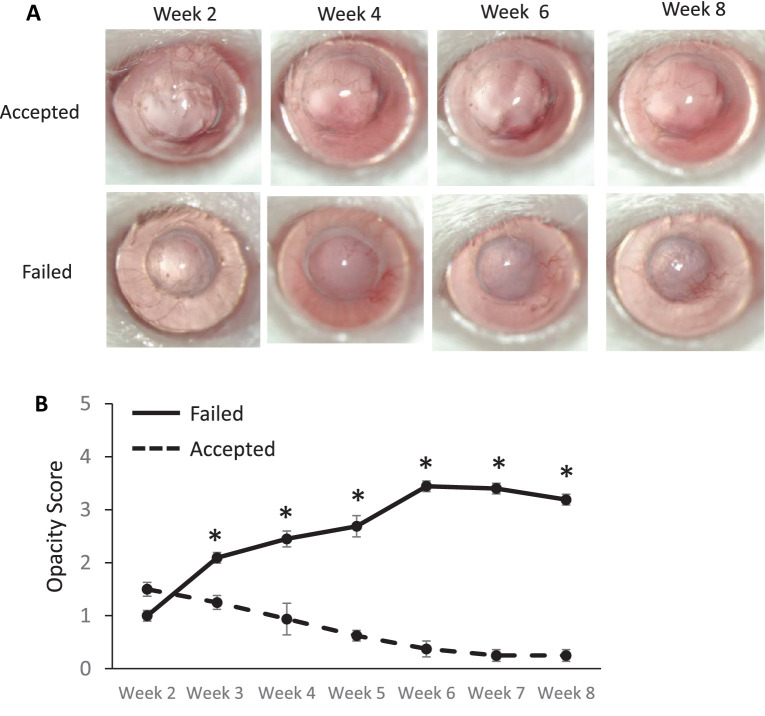
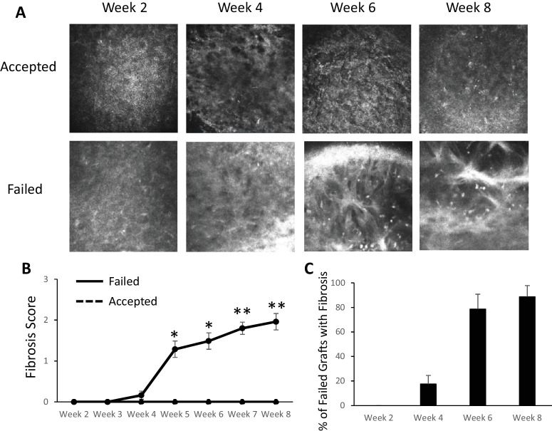
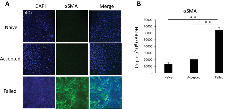
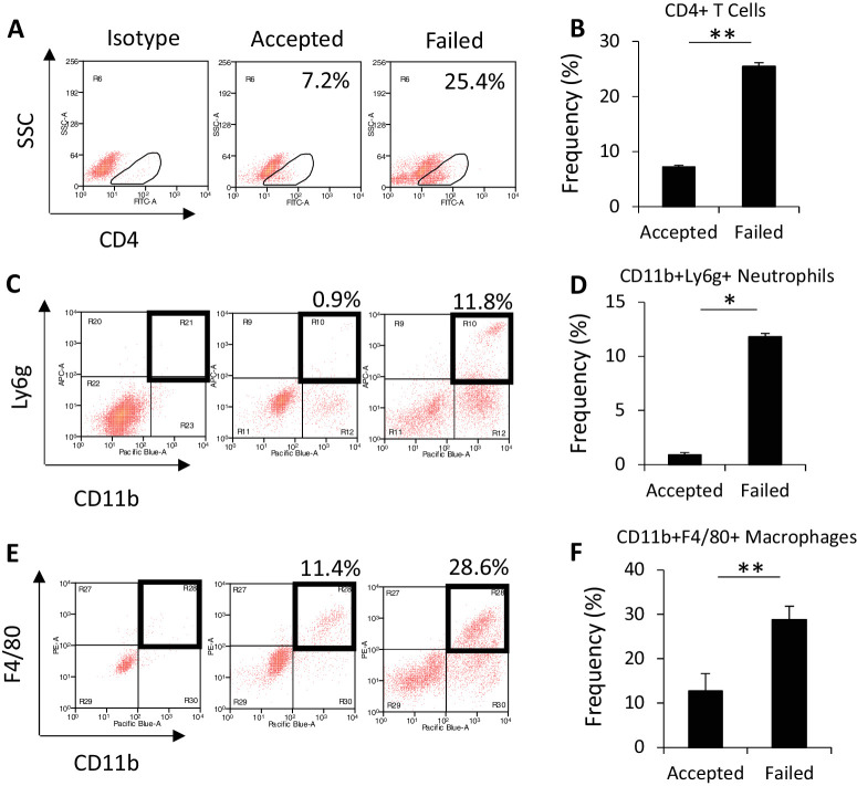
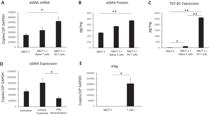
Methods: Allogeneic corneal transplantation was performed in mice. Graft opacity was evaluated by slit-lamp biomicroscopy, and fibrosis was assessed by in vivo confocal microscopy. Expression of alpha-smooth muscle actin (α-SMA) in both accepted and failed grafts was assessed by real-time PCR and immunohistochemistry. Frequencies of graft-infiltrating CD4+ T cells, neutrophils, and macrophages were assessed using flow cytometry. In vitro, MK/T-1 corneal fibroblasts were co-cultured with activated CD4+CD25- effector T cells isolated from corneal transplant recipient mice, and α-SMA expression was quantified by real-time PCR and ELISA. Neutralizing antibody was used to evaluate the role of interferon gamma (IFN-γ) in promoting α-SMA expression.

Results: The majority of failed grafts demonstrated clinical signs of fibrosis which became most evident at week 6 after corneal transplantation. Failed grafts showed higher expression of α-SMA as compared to accepted grafts. Flow cytometry analysis showed a significant increase in CD4+ T cells in failed grafts compared to accepted grafts. Co-culture of activated CD4+CD25- effector T cells with corneal fibroblasts led to an increase in α-SMA expression by fibroblasts. Inhibition of IFN-γ in culture significantly suppressed this increase in α-SMA expression as compared to immunoglobulin G control.

Conclusions: Fibrosis contributes to graft opacity in corneal transplant failure and is mediated at least in part by effector CD4+ T cells via IFN-γ.


Conflict of interest statement

Disclosure: S. Wang, None; S.K. Mittal, None; S. Lee, None; A.E. Herrera, None; M. Krauthammer, None; E. Elbasiony, None; T. Blanco, None; H. Alemi, None; H. Nakagawa, None; S.K. Chauhan, None; R. Dana, None; T.H. Dohlman, None

Figures

Figure 1.
Figure 1.
Corneal opacity in accepted or failed allogeneic transplantation. Corneal grafts were harvested from C57BL/6 donors and transplanted to BALB/c recipients, and graft opacity was assessed weekly for 8 weeks. (A) Representative slit-lamp images showing accepted (top row) and failed (bottom row) corneas post-transplantation in vivo. (B) Accepted grafts (dashed line) remained clear, but grafts that failed developed graft opacity (solid line). *P < 0.05 (n = 20/group).
Figure 2.
Figure 2.
In vivo confocal microscopy evaluation of fibrosis in corneal transplantation. (A) Representative in vivo confocal microscopy images showing the development of hyperreflective, thickened bands in the anterior stroma in failed grafts (bottom row). (B) Fibrosis score increased in failed grafts (solid line) over time, but accepted grafts continued to have a corneal opacity score of 0 (dashed line). (C) The majority of failed grafts demonstrated signs of fibrosis on confocal microscopy, and fibrosis became most evident around week 6. *P < 0.05, **P < 0.005 (failed, n = 16; accepted, n = 14).
Figure 3.
Figure 3.
α-SMA expression by grafts 6 weeks after corneal transplantation. (A) Representative images showing immunohistochemistry staining of α-SMA in the graft center. Blue indicates DAPI (nuclei staining), and green indicates α-SMA (magnification, 40×). (B) mRNA levels of α-SMA determined by PCR. **P < 0.01 (n = 3/group; representative data from one of two repeats are shown).
Figure 4.
Figure 4.
Immune cell frequency in corneal transplantation. (A, C, E) Corneas were harvested at week 6 after corneal transplantation for flow cytometry. Representative images show CD4+ T cell, neutrophil, and macrophage populations in accepted or failed grafts, gated on CD45+ cells. (B, D, F) Frequency of CD4+ T cells, neutrophils, and macrophages. *P < 0.05, **P < 0.005 (n = 6/group; representative data from one of two repeats are shown).
Figure 5.
Figure 5.
Expression of α-SMA in corneal fibroblasts co-cultured with effector T cells. (A, B) CD4+CD25 effector T cells were sorted from corneal transplant recipient (allo-primed) and naive (unprimed) mice. MK/T-1 corneal fibroblasts demonstrated increased α-SMA mRNA and protein expression when co-cultured with activated CD4+CD25 effector T cells isolated from corneal transplant recipient mice 2 weeks post-transplantation. (C) TGF-β1 protein levels in the culture supernatant were significantly increased in the MK/T-1 with penetrating keratoplasty (corneal transplantation) (PK) effector T-cell group. Anti-CD3 and anti-CD28 antibodies were included in all groups. Naïve T cells, effector T cells from naïve mice; PK T cells, effector T cells from corneal transplant recipient mice. (D) When MK/T-1 corneal fibroblasts were co-cultured with effector T cells from corneal transplant mice, inhibition of IFN-γ with neutralizing antibody suppressed α-SMA expression compared to IgG control. (E) The expression of IFN-γ mRNA by T cells was significantly greater than IFN-γ expression by fibroblasts. *P < 0.05, **P < 0.005 (n = 6/group; representative data from one of two repeats are shown).

References

    1. Coco G, Romano V. Corneal disease & transplantation. J Clin Med. 2022; 11: 4432. - PMC - PubMed
    1. Dana MR, Qian Y, Hamrah P. Twenty-five–year panorama of corneal immunology. Cornea. 2000; 19: 625–643. - PubMed
    1. Hamrah P. High-risk penetrating keratoplasty. Arch Soc Esp Oftalmol. 2005; 80: 5–7. - PubMed
    1. EBAA. 2014 Eye Banking Statistical Report. Washington, DC: Eye Bank Association of America; 2014.
    1. Coster DJ, Williams KA. The impact of corneal allograft rejection on the long-term outcome of corneal transplantation. Am J Ophthalmol. 2005; 140: 1112–1122. - PubMed