

SPLIT: Statistical Positronium Lifetime Image Reconstruction via Time-Thresholding
- PMID: 38261489
- PMCID: PMC11409919
- DOI: 10.1109/TMI.2024.3357659
SPLIT: Statistical Positronium Lifetime Image Reconstruction via Time-Thresholding
Abstract
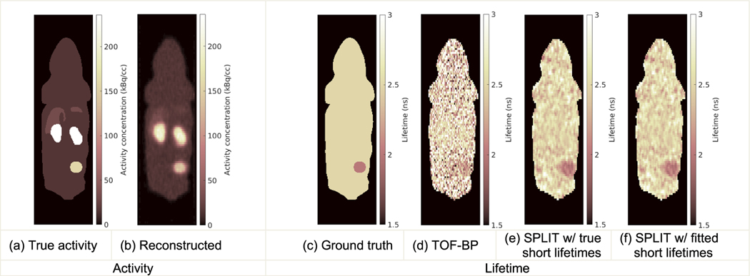
Positron emission tomography (PET) is a widely utilized medical imaging modality that uses positron-emitting radiotracers to visualize biochemical processes in a living body. The spatiotemporal distribution of a radiotracer is estimated by detecting the coincidence photon pairs generated through positron annihilations. In human tissue, about 40% of the positrons form positroniums prior to the annihilation. The lifetime of these positroniums is influenced by the microenvironment in the tissue and could provide valuable information for better understanding of disease progression and treatment response. Currently, there are few methods available for reconstructing high-resolution lifetime images in practical applications. This paper presents an efficient statistical image reconstruction method for positronium lifetime imaging (PLI). We also analyze the random triple-coincidence events in PLI and propose a correction method for random events, which is essential for real applications. Both simulation and experimental studies demonstrate that the proposed method can produce lifetime images with high numerical accuracy, low variance, and resolution comparable to that of the activity images generated by a PET scanner with currently available time-of-flight resolution.
Figures


Similar articles
-
High-resolution positronium lifetime tomography by the method of moments.Phys Med Biol. 2024 Dec 4;69(24):10.1088/1361-6560/ad9543. doi: 10.1088/1361-6560/ad9543. Phys Med Biol. 2024. PMID: 39569940
-
Positronium Lifetime Image Reconstruction for TOF PET.IEEE Trans Med Imaging. 2022 Oct;41(10):2848-2855. doi: 10.1109/TMI.2022.3174561. Epub 2022 Sep 30. IEEE Trans Med Imaging. 2022. PMID: 35584079 Free PMC article.
-
Fast high-resolution lifetime image reconstruction for positron lifetime tomography.Commun Phys. 2025;8(1):181. doi: 10.1038/s42005-025-02100-6. Epub 2025 Apr 26. Commun Phys. 2025. PMID: 40291544 Free PMC article.
-
Performance assessment of the 2 γpositronium imaging with the total-body PET scanners.EJNMMI Phys. 2020 Jun 30;7(1):44. doi: 10.1186/s40658-020-00307-w. EJNMMI Phys. 2020. PMID: 32607664 Free PMC article.
-
Magnetic resonance-guided positron emission tomography image reconstruction.Semin Nucl Med. 2013 Jan;43(1):30-44. doi: 10.1053/j.semnuclmed.2012.08.006. Semin Nucl Med. 2013. PMID: 23178087 Free PMC article. Review.
Cited by
-
Phantom imaging demonstration of positronium lifetime with a long axial field-of-view PET/CT and 124I.EJNMMI Phys. 2025 Aug 26;12(1):80. doi: 10.1186/s40658-025-00790-z. EJNMMI Phys. 2025. PMID: 40855031 Free PMC article.
-
Positronium image of the human brain in vivo.Sci Adv. 2024 Sep 13;10(37):eadp2840. doi: 10.1126/sciadv.adp2840. Epub 2024 Sep 13. Sci Adv. 2024. PMID: 39270027 Free PMC article.
-
High-Resolution Positronium Lifetime Tomography at Clinical Activity Levels on the PennPET Explorer.J Nucl Med. 2025 Sep 2;66(9):1464-1470. doi: 10.2967/jnumed.125.270130. J Nucl Med. 2025. PMID: 40841149 Free PMC article.
-
Positronium lifetime validation measurements using a long-axial field-of-view positron emission tomography scanner.EJNMMI Phys. 2024 Aug 30;11(1):76. doi: 10.1186/s40658-024-00678-4. EJNMMI Phys. 2024. PMID: 39210079 Free PMC article.
-
High-resolution positronium lifetime tomography by the method of moments.Phys Med Biol. 2024 Dec 4;69(24):10.1088/1361-6560/ad9543. doi: 10.1088/1361-6560/ad9543. Phys Med Biol. 2024. PMID: 39569940
References
-
- Beyer T et al., “A combined PET/CT scanner for clinical oncology,” J. Nucl. Med, vol. 41, no. 8, pp. 1369–1379, Aug. 2000. - PubMed
Publication types
MeSH terms
Grants and funding
LinkOut - more resources
Full Text Sources
Medical
Miscellaneous
