. 2024 Feb 27;43(2):113685.
doi: 10.1016/j.celrep.2024.113685. Epub 2024 Jan 22.

Compartment-specific regulation of NaV1.7 in sensory neurons after acute exposure to TNF-α

Affiliations

Compartment-specific regulation of NaV1.7 in sensory neurons after acute exposure to TNF-α

Sidharth Tyagi et al. Cell Rep. .

Abstract

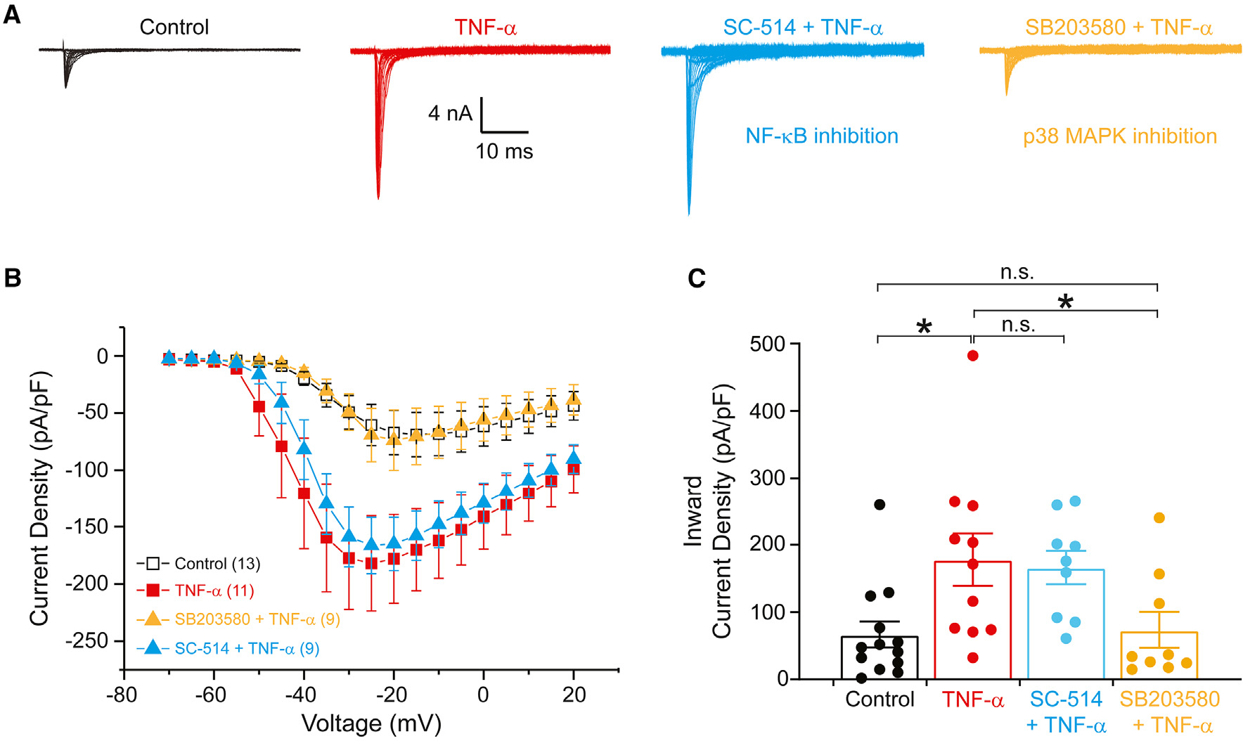
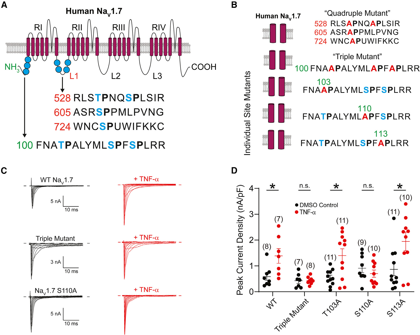
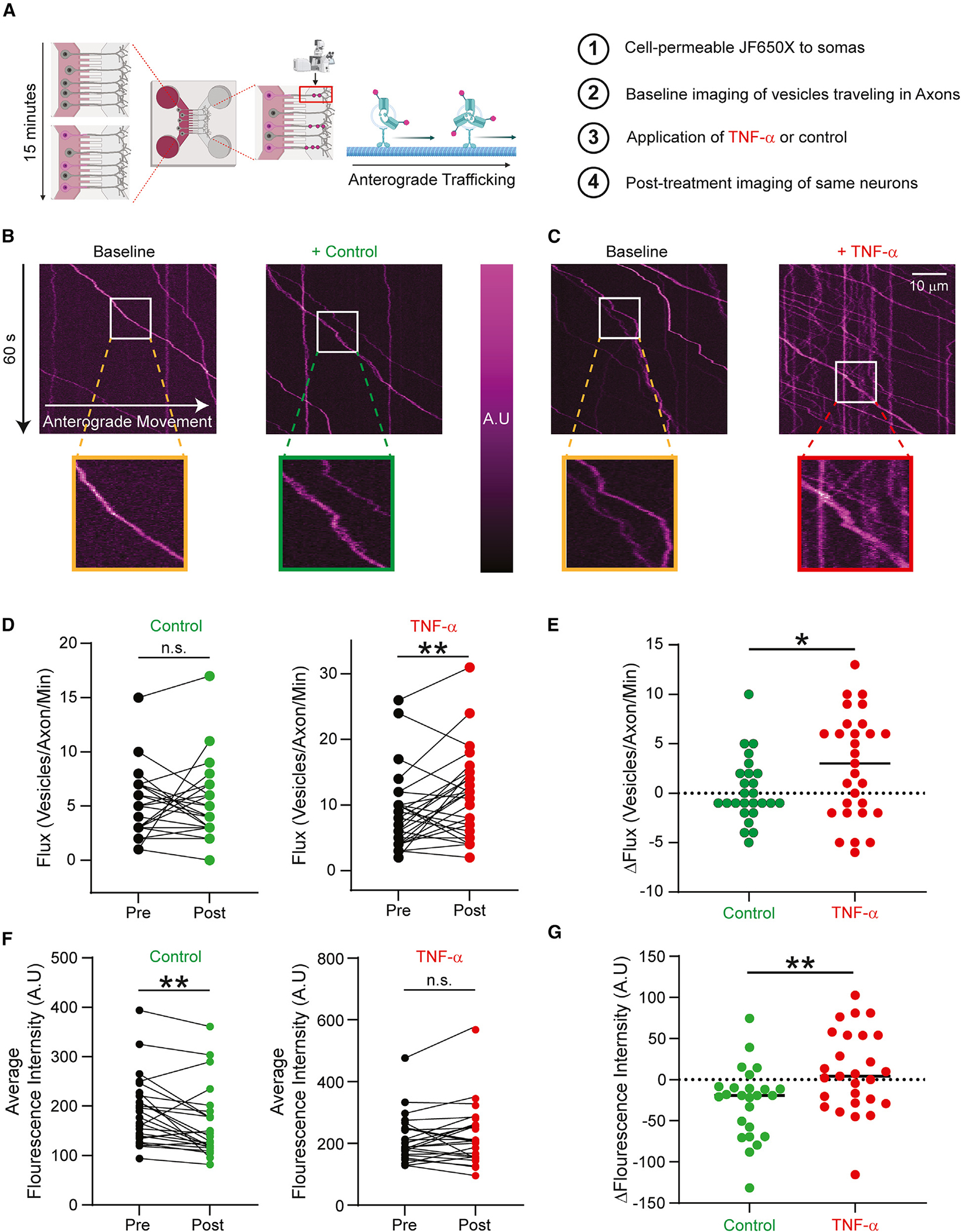
Tumor necrosis factor α (TNF-α) is a major pro-inflammatory cytokine, important in many diseases, that sensitizes nociceptors through its action on a variety of ion channels, including voltage-gated sodium (NaV) channels. We show here that TNF-α acutely upregulates sensory neuron excitability and current density of threshold channel NaV1.7. Using electrophysiological recordings and live imaging, we demonstrate that this effect on NaV1.7 is mediated by p38 MAPK and identify serine 110 in the channel's N terminus as the phospho-acceptor site, which triggers NaV1.7 channel insertion into the somatic membrane. We also show that the N terminus of NaV1.7 is sufficient to mediate this effect. Although acute TNF-α treatment increases NaV1.7-carrying vesicle accumulation at axonal endings, we did not observe increased channel insertion into the axonal membrane. These results identify molecular determinants of TNF-α-mediated regulation of NaV1.7 in sensory neurons and demonstrate compartment-specific effects of TNF-α on channel insertion in the neuronal plasma membrane.

Keywords: CP: Neuroscience; Na(V)1.7; TNF-α; distal axons; inflammatory pain; neuronal compartments; soma.


Conflict of interest statement

Declaration of interests The authors declare no competing interests.

Figures

Figure 1.
Figure 1.. Native TTX-S and TTX-R neuronal sodium currents, as well as human NaV1.7 currents, are increased by a short application of TNF-α
(A) Representative TTX-S and TTX-R current traces recorded from DRG neurons in the absence (left) and presence (right) of 20 ng/mL TNF-α for 20 min (B) Current density-voltage relationships for TTX-S and TTX-R currents in the absence or presence of TNF-α. (C) Inward current density through TTX-S and TTX-R sodium channels both increased in the presence of TNF-α (n = 21) vs. vehicle (n = 22). *p < 0.05 by unpaired Student’s t test. (D) Conductance-voltage relationships of TTX-S and TTX-R sodium currents recorded from DRG neurons. Application of TNF-α hyperpolarizes the voltage dependence of activation of both TTX-S and TTX-R sodium channels. Conductance-voltage (G-V) relationships were fit by Boltzmann equations. The midpoint (V1/2) and slope (k) values for the voltage-dependent activation curves of TTX-S channels were −28.27 ± 1.13 and 5.02 ± 0.21 mV in the absence of TNF-α and −33.50 ± 1.37 and 4.21 ± 0.29 mV in the presence of TNF-α, respectively. The V1/2 and k values for the voltage-dependent activation curves of TTX-R channels were −19.86 ± 0.90 and 5.02 ± 0.14 mV in the absence of TNF-α and −23.50 ± 1.19 and 5.02 ± 0.17 mV in the presence of TNF-α, respectively. (E) Representative hNaV1.7 current traces recorded from DRG neurons in the absence (left) and presence (right) of 20 ng/mL TNF-α for 20 min. Endogenous TTX-R currents were minimal. (F) Current density-voltage relationships for hNaV1.7 in the absence and presence of TNF-α. (G) Inward current density through hNaV1.7 increased in the presence of TNF-α (n = 13 untreated, 11 treated) vs. vehicle (n = 6 untreated, 6 treated). *p < 0.05 by unpaired Student’s t test. (H) Conductance-voltage relationships of hNaV1.7 sodium currents recorded from DRG neurons. G-V relationships were fit by Boltzmann equations. Application of TNF-α hyperpolarizes the voltage-dependence of activation of hNaV1.7. The V1/2 and k values for the voltage-dependent activation curves of neurons treated with DMSO control were −26.4 ± 1.1 and 6.2 ± 0.3 mV. The V1/2 and k values for the voltage-dependent activation curves of neurons treated with TNF-α were −32.5 ± 1.3 and 4.6 ± 0.5 mV. Data are presented as mean ± SEM.
Figure 2.
Figure 2.. Acute exposure to TNF-α causes hyperexcitability of DRG neurons
Current-clamp recordings of DRG neurons expressing hNaV1.7 treated with TNF-α (red) or vehicle (DMSO; black). (A) Action potentials elicited by injection of 200 pA current in DRG neurons expressing hNaV1.7 treated with TNF-α (right) vs. vehicle (left). (B) Action potentials elicited by injection of 500 pA current in DRG neurons expressing hNaV1.7 treated with TNF-α (right) vs. vehicle (left). (C) Quantification of repetitive firing of action potentials provoked by current injections of increasing amplitude. (D) Recordings from neurons subjected to 500 ms ramp current injection from Resting Membrane Potential (RMP) to 1 nA (2 pA/ms). (E) Recordings from neurons subjected to 500 ms ramp current injection from RMP to 2 nA (4 pA/ms). (F) Quantification of action potentials (APs) evoked by ramp current injection from 0.5 to 2 nA by 500 ms ramp current injections of four different speeds. (G) Latency of AP generation was quantified as the time required from stimulus onset to the peak of the first AP. AP latency of neurons treated with TNF-α was lower than those treated with vehicle. Data are presented as mean ± SEM. *p < 0.01 by unpaired Student’s t tests with Bonferroni correction for multiple comparisons.
Figure 3.
Figure 3.. Acute enhancement of hNaV1.7 current by TNF-α is dependent on the p38 MAPK
(A) Representative hNaV1.7 currents recorded from DRG neurons in the absence (black) and presence (red) of 20 ng/mL TNF-α for 20 min. Blue traces are from TNF-α-exposed neurons preincubated with 20 μM SC-514 (an inhibitor of NF-κB) for 1 h. Gold traces are from TNF-α-exposed neurons preincubated with 20 μM SB203580 (an inhibitor of the p38 MAPK) for 30 min (B) Current density-voltage relationships for hNaV1.7 sodium currents in the absence and presence of TNF-α and with preincubation with inhibitors of NF-κB or p38 MAPK. (C) Comparison of maximal hNaV1.7 current density between neurons treated with DMSO control (n = 13), TNF-α (n = 11), or TNF-α and inhibitors of NF-κB (SC-514, n = 9) and p38 MAPK (SB203580, n = 9). Data are presented as mean ± SEM. *p < 0.05 vs. control and SB203580 + TNF-α by one-way ANOVA followed by the Bonferroni post-hoc test. Data are presented as mean ± SEM.
Figure 4.
Figure 4.. The phosphorylation site S110 in the NaV1.7 N terminus is critical for acute upregulation of human NaV1.7 current by TNF-α
(A) Schematic depicting the membrane topology of human NaV1.7. Putative phosphorylation sites in the L1 (red numbers) and N terminus (green numbers) marked in blue. The schematic was adapted from Tyagi et al. with the permission of the authors. (B) Phosphorylation site mutants generated in this study. Putative phosphorylation sites are marked in blue, and alanine substitutions are depicted in red. (C) Representative NaV1.7 current traces in the absence (left) and presence (right) of TNF-α from NaV1.8-null mouse neurons expressing (from top to bottom) WT, triple mutant, and S110A NaV1.7 channels. (D) Peak current densities from NaV1.8-null neurons expressing NaV1.7 phosphorylation site mutants. Data are presented as mean ± SEM. * p < 0.05 by unpaired t test.
Figure 5.
Figure 5.. Acute exposure to TNF-α drives packaging of NaV1.7 and vesicular flux to the axons of DRG neurons
(A) Schematic of OPAL imaging. Cell-permeable JF650X-HaloTag is added to the somatic chamber of MFCs containing DRG neurons expressing Halo-NaV1.7. Time lapses of vesicular trafficking in axons were taken before and after perfusion of 20 ng/mL TNF-α or vehicle (imaging saline). (B) Representative kymographs from an axon containing multiple anterogradely trafficking vesicles carrying Halo-NaV1.7 before and 20 min after perfusion of vehicle. Insets show the signal intensity of the magnified tracks. (C) Representative kymographs from an axon containing multiple anterogradely trafficking vesicles carrying Halo-NaV1.7 before and 20 min after perfusion of TNF-α. Insets show the signal intensity of the magnified tracks. (D) Flux of vesicles carrying Halo-NaV1.7 significantly increased following acute application of TNF-α. Vehicle perfusion did not affect the flux of anterogradely moving Halo-NaV1.7-positive vesicles. **p < 0.01, paired Student’s t test. (E) Neurons exposed to TNF-α had significantly increased flux of vesicles carrying Halo-NaV1.7 compared to vehicle. *p < 0.05 by unpaired Student’s t test. (F) Quantitation of the average fluorescence intensity of anterogradely moving vesicles carrying Halo-NaV1.7. Fluorescence intensity of Halo-NaV1.7 vesicles was significantly lower following vehicle perfusion; intensity was not different following TNF-α application. **p < 0.01, paired Student’s t test. Scale bars: 10 μm (G) The change in fluorescence intensity of vesicles carrying Halo-NaV1.7 following TNF-α application was significantly higher than the one produced by vehicle. *p < 0.05, unpaired Student’s t test. Data are presented as mean ± SEM.
Figure 6.
Figure 6.. Insertion of NaV1.7 in response to acute TNF-α exposure is differentially regulated in somas and axons
(A) Schematic of insertion assay. DRG neurons plated in MFC chambers enabled the compartment-specific investigation of insertion dynamics. First, channels at the cell surface were labeled with cell-impermeable JF549i-HaloTag ligand. Excess ligand was thoroughly washed away, and the second cell-impermeable ligand (JF-635i) was added to the bath. Newly inserted channels would be labeled by fluorophore present in the bath and fluoresce in the far-red channel of the spinning-disk confocal microscope. (B) Representative images of DRG neuronal somas before (left) and after (right) exposure to TNF-α for 20 min. Top: somas of DRG neurons expressing WT Halo-NaV1.7. Bottom: somas from DRG neurons expressing Halo-NaV1.7 S110A. (C) Acute application of TNF-α drives insertion of WT Halo-NaV1.7 channels to the somatic membrane of DRG neurons above the baseline rate of insertion. Mutation of the S110 residue reduces this effect. (D) Representative images captured from the distal axons of DRG neurons before (left) and after (right) exposure to TNF-α for 20 min. Top: axons of DRG neurons expressing WT Halo-NaV1.7. Bottom: axons of DRG neurons expressing Halo-NaV1.7 S110A. Because only newly inserted channels were labeled with Halo-JF635i ligand, the baseline fluorescence intensity of distal axons was very low in the far-red wavelength. Representative images have been background subtracted so that the distal axonal signal can be better visualized by readers. The image adjustment was identical for both vehicle- and TNF-α-treated conditions. (E) TNF-α does not affect the insertion kinetics of WT Halo-NaV1.7 channels at the distal axons of DRG neurons. There were no statistically significant differences between the insertion of WT channels vs. those channels carrying the S110A mutation. (F) Representative images captured from ND7/23 cells transfected with hNaV1.7-NTHalo before (left) and after (right) exposure to vehicle or 20 ng/mL TNF-α for 20 min. (G) Acute application of TNF-α drives insertion of hNaV1.7-NTHalo to the somatic membrane relative to vehicle. Data from each cell are normalized to the fluorescence intensity at baseline (preperfusion). *p < 0.05 and **p < 0.01 by unpaired Student’s t test. n.s. (not significant), p > 0.05 by unpaired Student’s t test. Scale bars: 10 μm. Data are presented as mean ± SEM.
Figure 7.
Figure 7.. Summary of the acute effect of TNF-α on DRG neurons
In response to tissue damage, macrophages release TNF-α in the DRG. Acutely, TNF-α binding to a TNFR initiates a phosphorylation cascade. The p38 MAPK is phosphorylated, which subsequently phosphorylates NaV1.7 at the N-terminal site S110. This leads to an increase in insertion kinetics of the channel at the somatic membrane. Increased somatic NaV1.7 could lead to increased ectopic discharge, enhanced peripheral AP propagation, and soma-to-soma sensitization.

Similar articles

Cited by

References

    1. Amir R, Argoff CE, Bennett GJ, Cummins TR, Durieux ME, Gerner P, Gold MS, Porreca F, and Strichartz GR (2006). The Role of Sodium Channels in Chronic Inflammatory and Neuropathic Pain. J. Pain 7. S1–29. - PubMed
    1. Kidd BL, and Urban LA (2001). Mechanisms of inflammatory pain. Br. J. Anaesth. 87, 3–11. - PubMed
    1. Talbot S, Foster SL, and Woolf CJ (2016). Neuroimmunity: Physiology and Pathology. Annu. Rev. Immunol. 34, 421–447. - PubMed
    1. Bennett DL, Clark AJ, Huang J, Waxman SG, and Dib-Hajj SD (2019). The Role of Voltage-Gated Sodium Channels in Pain Signaling. Physiol. Rev. 99, 1079–1151. - PubMed
    1. Dib-Hajj SD, Yang Y, Black JA, and Waxman SG (2013). The NaV1.7 sodium channel: from molecule to man. Nat. Rev. Neurosci. 14, 49–62. - PubMed

Publication types

MeSH terms

Substances

LinkOut - more resources