. 2024 Jan 30;121(5):e2311487121.
doi: 10.1073/pnas.2311487121. Epub 2024 Jan 23.

Tracking the role of Aire in immune tolerance to the eye with a TCR transgenic mouse model

Affiliations

Tracking the role of Aire in immune tolerance to the eye with a TCR transgenic mouse model

Mianmian Yin et al. Proc Natl Acad Sci U S A. .

Abstract

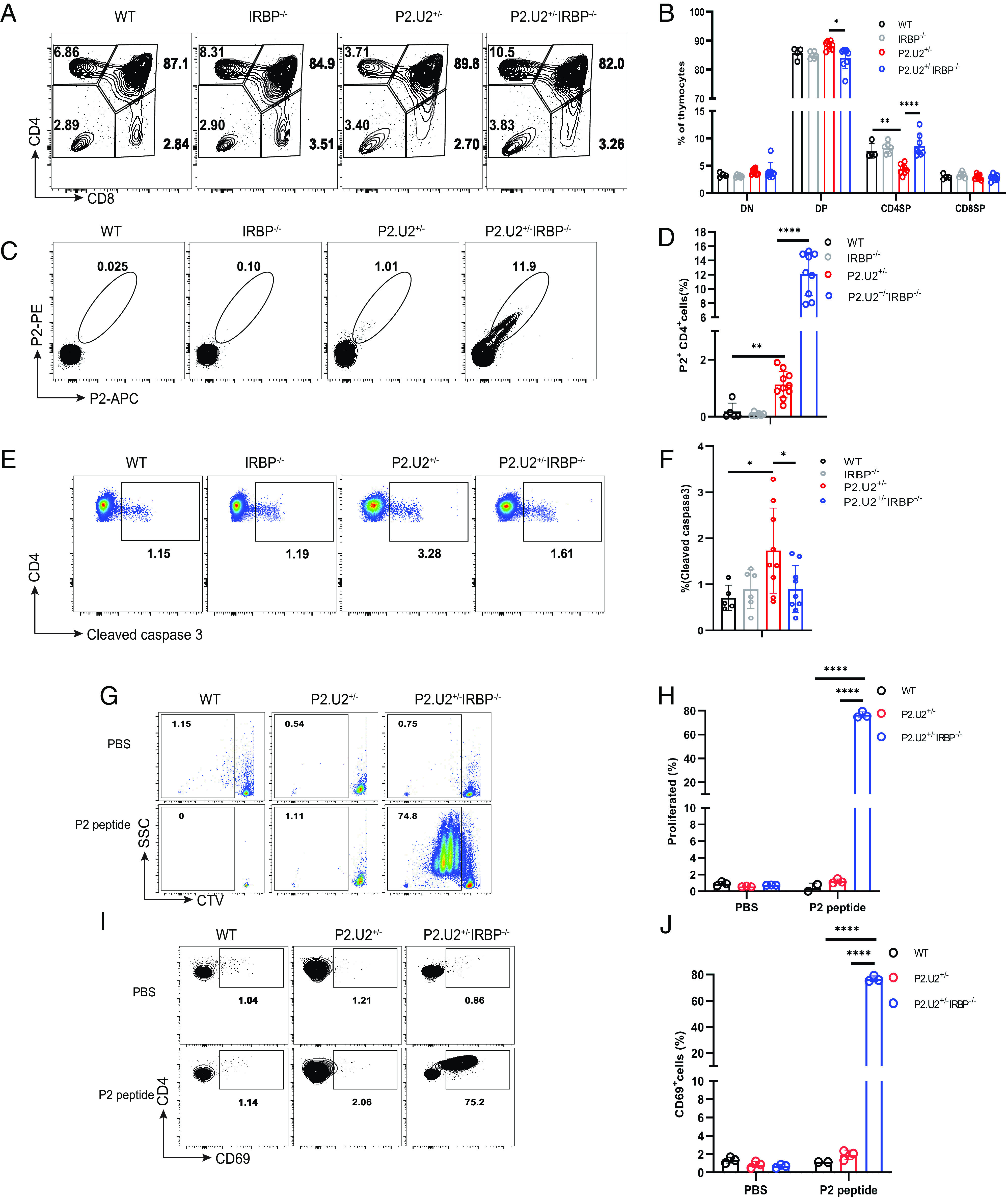
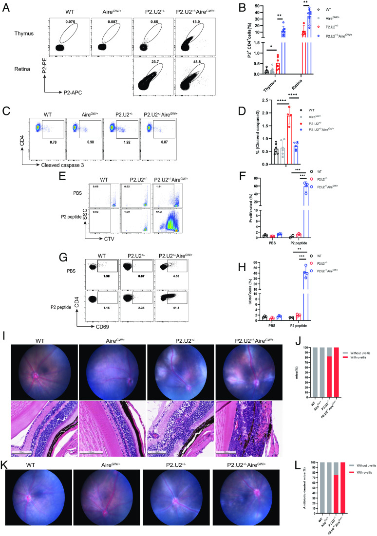
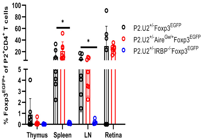
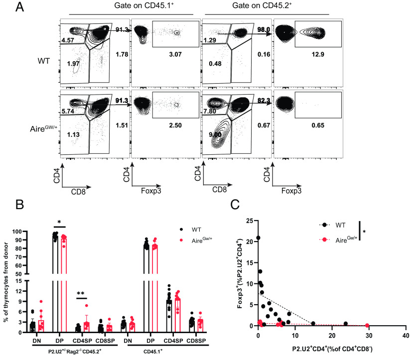
Roughly one-half of mice with partial defects in two immune tolerance pathways (AireGW/+Lyn-/- mice) spontaneously develop severe damage to their retinas due to T cell reactivity to Aire-regulated interphotoreceptor retinoid-binding protein (IRBP). Single-cell T cell receptor (TCR) sequencing of CD4+ T cells specific for a predominate epitope of IRBP showed a remarkable diversity of autoantigen-specific TCRs with greater clonal expansions in mice with disease. TCR transgenic mice made with an expanded IRBP-specific TCR (P2.U2) of intermediate affinity exhibited strong but incomplete negative selection of thymocytes. This negative selection was absent in IRBP-/- mice and greatly defective in AireGW/+ mice. Most P2.U2+/- mice and all P2.U.2+/-AireGW/+ mice rapidly developed inflammation of the retina and adjacent uvea (uveitis). Aire-dependent IRBP expression in the thymus also promoted Treg differentiation, but the niche for this fate determination was small, suggesting differences in antigen presentation leading to negative selection vs. thymic Treg differentiation and a stronger role for negative selection in preventing autoimmune disease in the retina.

Keywords: Aire; IRBP; autoimmune; negative selection; uveitis.


Conflict of interest statement

Competing interests statement:The authors declare no competing interest.

Figures

Fig. 1.
Fig. 1.
AireGW/+Lyn−/− mice do not develop uveitis in the absence of IRBP. (A) Representative funduscopic images of 3-mo-old WT mice without uveitis, AireGW/+Lyn−/−IRBP+/+ mice with uveitis, AireGW/+Lyn−/−IRBP+/– mice with uveitis, and AireGW/+Lyn−/−IRBP−/− mice without uveitis. (B) Frequencies of AireGW/+Lyn−/−IRBP+/+ (n = 8), AireGW/+Lyn−/−IRBP+/– (n = 10) and AireGW/+Lyn−/−IRBP−/− (n = 22) mice with uveitis and without uveitis. Fisher’s exact test, AireGW/+Lyn−/−IRBP+/+ vs. AireGW/+Lyn−/−IRBP−/−, P = 0.0138; AireGW/+Lyn−/−IRBP+/– vs. AireGW/+Lyn−/−IRBP−/−, P = 0.0013.
Fig. 2.
Fig. 2.
TCR clonotypes of P2/I-Ab tetramer-binding CD4+ T cells from LN of AireGW/+Lyn−/− mice. (A) Experimental schematic. Single-cell suspensions of LN (pooled from eye-draining cervical, submandibular, and axillary lymph nodes) from AireGW/+Lyn−/− mice with or without uveitis were incubated with IRBP-P2 tetramer. P2 tetramer-binding CD4+T (P2+CD4+ T) cells were isolated by magnetic beads enrichment followed by cell sorting and single-cell TCRα and β sequencing. (B) Size distributions of TCR clonotypes of P2+CD4+ T cells in LN of AireGW/+Lyn−/− mice with and without uveitis at 9 wk of age, representing 416 cells in two datasets from seven pooled mice and eight pooled mice, and 380 cells in one dataset from 16 pooled mice, respectively. Cells of the same TCR clonotype are shown as a single pie slice representing the percent of these cells in the entire sample. (C) Gini index for TCR clonotypes in the P2+CD4+T cells in LN of AireGW/+Lyn−/− mice with and without uveitis. (D) Chain pairing of TCRVα and TCRVβ from P2+CD4+ T cells in LN of AireGW/+Lyn−/− mice with and without uveitis are displayed as chord diagrams, where the thickness of ribbons connecting chains indicates the frequency of pairing. (E) The V, D, and J segment usage and CDR3 sequences of top five most expanded TCR clonotypes in P2+CD4+ T cells from AireGW/+Lyn−/− mice with uveitis in dataset 1. n.i.: not identified. (F) Representative flow cytometric analysis of NFAT-GFP induction in hybridoma cells expressing empty vector (EV) or TCR clonotypes (1, 2, 3, 4, and 5) after stimulation with P2 tetramer for 24 h. (G) Frequency of GFP in (F). (H) Representative flow cytometric analysis of GFP in hybridomas cells expressing empty vector (EV) or TCRs clonotypes (1, 2, 3, 4, and 5) after stimulation with different doses of P2 peptide in the presence of DC of Lyn−/− mice for 24 h. (I) Frequency of GFP in (H). **P < 0.01, ***P < 0.001, and ****P < 0.0001. Two-tailed t test; error bars are the mean ± SD (n = 3).
Fig. 3.
Fig. 3.
P2.U2+/− transgenic mice spontaneously develop uveitis. (A) Representative flow cytometric analysis of CD4 and CD8 in the thymus, spleen and LN of WT (n = 6) or P2.U2+/− (n = 6) mice at 5 to 7 wk, gated on live lymphocytes. (B) Frequencies (Left) and cell number (Right) of CD4CD8(DN), CD4+CD8+(DP), CD4+CD8(CD4SP) and CD4CD8+(CD8SP) populations of thymus in panel (A). Data were pooled from at least three independent experiments. (C) Frequencies of CD4SP and CD8SP populations of spleen and LN in panel (A). (D) CD4+CD8/CD4CD8+ ratios in the thymus, spleen, and LN in panel (B) and (C). (E) Representative funduscopic images of 5 to 7 wk old WT mice without uveitis or P2.U2+/− mice with uveitis. (F) Frequencies of WT (n = 23) or P2.U2+/− (n = 33) mice with uveitis and without uveitis at 5 to 7 wk. Data are pooled from at least four independent experiments. Fisher’s exact test, P2.U2+/− vs. WT, P < 0.0001. (G) Representative flow cytometric analysis of P2+CD4+ T cells in the thymus of WT mice (n = 5) and in the thymus and retina of P2.U2+/− mice (thymus: n = 5; retina n = 6), gated on the TCRβ+CD4+CD8DUMP cells. Data were pooled from at least three independent experiments. (H) Frequencies of P2+CD4+ in the thymus or retina in panel (G). *P < 0.05; **P < 0.01; ***P < 0.001; ****P < 0.0001. Two-tailed t test; error bars are mean ± SD.
Fig. 4.
Fig. 4.
P2.U2 TCR enhances negative selection of CD4SP thymocytes in an IRBP-dependent manner. (A and B) Representative flow cytometric analysis (A) and frequencies (B) of T cell subtypes in the thymus of WT (n = 4), IRBP−/− (n = 6), P2.U2+/− (n = 8) and P2.U2+/−IRBP−/− (n = 8) mice at 5 to 7 wk of age. (C and D) Representative flow cytometric analysis (C) and frequencies (D) of P2+ binding by CD4SP thymocytes of WT (n = 5), IRBP−/− (n = 6), P2.U2+/− (n = 10), and P2.U2+/−IRBP−/− (n = 9) mice at 5 to 7 wk of age. Cells shown were gated on the TCRβ+CD4+CD8DUMP cells. (E and F) Representative flow cytometric analysis (E) and frequencies (F) of the presence of cleaved caspase 3 in CD4SP thymocytes of WT (n = 5), IRBP−/− (n = 6), P2.U2+/− (n = 9) and P2.U2+/−IRBP−/− (n = 9) mice at 5 to 7 wk. (GJ) CTV-labeled CD4+ T cells from spleens of WT, P2.U2+/−, or P2.U2+/−IRBP−/−mice were cultured in vitro in the presence of PBS or 100 ng/mL P2 peptide with CD11c+ dendritic cells (DCs) isolated from the spleen of WT mice. Dilution of CTV and expression of CD69 was assessed by flow cytometry on day 3. (G and H) Representative flow cytometric analysis (G) and frequencies (H) of dilution of CTV in CTV-labeled CD4+ T cells from WT (PBS: n = 3; P2 peptide: n = 2), P2.U2+/− (n = 3) or P2.U2+/−IRBP−/− (n = 3) mice incubated in vitro with PBS or P2 peptide. (I and J) Representative flow cytometric analysis (I) and frequencies (J) of expression of CD69 on CTV-labeled CD4+ T cells of WT (PBS: n = 3; P2 peptide: n = 2), P2.U2+/− (n = 3) or P2.U2+/−IRBP−/− (n = 3) mice stimulated with PBS or P2 peptide. Data were pooled from at least three independent experiments (AF) or two independent experiments (GJ). In (B, F, H, and J), one-way ANOVA with Tukey’s multiple comparisons tests were used. In (D), two-tailed t tests were used. *P < 0.05; **P < 0.01; ****P < 0.0001; error bars are mean ± SD.
Fig. 5.
Fig. 5.
AireGW/+ mice have greatly reduced negative selection of P2.U2 TCR transgenic T cells. (A and B) Representative flow cytometric analysis (A) and frequencies (B) of P2+CD4+ T cells in the thymus, gated on TCRβ+CD4+CD8DUMPthymocytes, and retina, gated on TCRβ+CD4+CD8DUMPT cells, of WT (n = 4), AireGW/+ (n = 8), P2.U2+/− (thymus: n = 6; retina: n = 4) and P2.U2+/−AireGW/+ (n = 8) mice at 5 to 7 wk of age. Data were pooled from at least three independent experiments. For thymus data of the P2.U2+/− group of panel (B), two of the mice were also included in the data shown in (Fig. 3H). (C and D) Representative flow cytometric analysis (C) and frequencies (D) of cleaved caspase 3 staining in CD4+CD8- thymocytes of WT (n = 4), AireGW/+ (n = 6), P2.U2+/− (n = 4) and P2.U2+/−AireGW/+ (n = 6) mice at 5 to 7 wk. Data were pooled from at least three independent experiments. (EH) CTV-labeled splenic CD4+ T cells from WT, P2.U2+/−, or P2.U2+/−AireGW/+ mice were cultured in vitro with CD11c+ DCs, isolated from the spleens of WT mice, in the presence of PBS or 100 ng/mL P2 peptide. Dilution of CTV and expression of CD69 was assessed by flow cytometry on day 3. Representative flow cytometric analysis (E and G) and frequencies (F and H) of dilution of CTV (E and F) and CD69 induction (G and H) in CTV-labeled splenic CD4+ T cells from WT (n = 3), P2.U2+/− (n = 3) or P2.U2+/−AireGW/+ (n = 4) mice. Data were pooled from two independent experiments. For the data of the WT and P2.U2+/− groups of panels (F and H), the data is the same for these groups as shown in Fig. 4 H and J. (I) Representative funduscopic images (Top row) or H&E-stained retinal sections (Bottom row) for 2- to 4-mo-old WT, AireGW/+ mice without uveitis, and P2.U2+/−, P2.U2+/−AireGW/+ mice with uveitis. (J) Frequencies of uveitis or lack of uveitis in WT (n = 23), AireGW/+ (n = 16), P2.U2+/− (n = 33) and P2.U2+/−AireGW/+ (n = 37) mice at 5 to 7 wk. Fisher’s exact test, P2.U2+/− vs. WT, P < 0.0001; P2.U2+/−AireGW/+vs. WT, P < 0.0001; P2.U2+/− vs. P2.U2+/−AireGW/+, P = 0.0084. For WT and P2.U2+/− group of Fig. 5J, the data is the same as the WT and P2.U2+/− groups of Fig. 3F. (K and L) Representative funduscopic images (K) and frequencies (L) of WT (n = 3), AireGW/+ (n = 16), P2.U2+/− (n = 12) and P2.U2+/−AireGW/+ (n = 4) mice with uveitis or without uveitis after antibiotic treatment for 6 to 9 wk starting at birth. Fisher’s exact test, P2.U2+/− vs. WT, P = 0.044; P2.U2+/−AireGW/+ vs. WT, P = 0.0286. In (B), two-tailed t tests were used. In (D, F, and H), one-way ANOVA with Tukey’s multiple comparisons tests were used. *P < 0.05; **P < 0.01; ***P < 0.001; ****P < 0.0001; error bars are mean ± SD.
Fig. 6.
Fig. 6.
A minority of P2.U2 TCR transgenic T cells specific for P2 tetramer adopt a Treg fate in the thymus of mice that express IRBP, but are enriched in the periphery. Frequencies of Foxp3EGFP+ of P2+ CD4+ T cells in the thymus, spleen, LN and retina of P2.U2+/−Foxp3EGFP (n = 7), P2.U2+/−AireGW/+Foxp3EGFP (n = 7) and P2.U2+/−IRBP−/−Foxp3EGFP (n = 5) mice at 6 to 7 wk of age. Cells shown were gated on TCRβ+CD4+CD8DUMPP2+ cells. Data were pooled from at least three independent experiments. Two-tailed t test was used, *P < 0.05; error bars are mean ± SD.
Fig. 7.
Fig. 7.
A small niche for thymic development of IRBP P2-specific Tregs is present in Aire-expressing mice. Mixed bone marrow chimeric mice were made in which bone marrow cells of P2.U2+/−Rag2−/−CD45.2+ male donor mice were engrafted along with various fractions of non-transgenic bone marrow cells from male CD45.1+ mice. Recipient mice were irradiated WT or AireGW/+CD45.1+/CD45.2+ mice. Six weeks post-engraftment, the fate of P2.U2+ T cells was analyzed. (A) Representative flow cytometric analysis of CD45.1+polyclonal T cells (Left) and P2.U2+Rag2−/−CD45.2+ T cells (Right) from recipient mice of the indicated Aire genotype receiving a small percentage of CD45.2+ bone marrow (WT recipient:1.93% CD45.2+ cells; AireGW/+:0.43% CD45.2+ cells). The left column (CD4 vs. CD8) depicts all cells in the lymphocyte gate; the right column (intracellular Foxp3 vs. CD4) depicts CD4+CD8 gated samples. (B) Frequencies of DN, DP, CD4SP and CD8SP populations of thymocytes for donor cells (P2.U2+Rag2−/−CD45.2+ cells and CD45.1+ cells) in WT (n = 16) or AireGW/+CD45.1+/CD45.2+ (n = 8) recipient mice in panel (A). (C) Summary plot of the efficiency of P2.U2+ Treg development, in which the frequencies of P2.U2+/−Rag2−/−CD45.2+ cells that express Foxp3 is plotted vs. the frequency of P2.U2+/−Rag2−/−CD45.2+CD4+ thymocytes in all donor CD4+CD8 cells isolated from recipient mice of the indicated genotype. Dashed lines indicate simple linear regression line [WT (n = 17): R = 0.298; AireGW/+ (n = 9): R = 0.249]. *P < 0.05; **P < 0.01. In (B and C), two-tailed t test; error bars are mean ± SD.

Similar articles

Cited by

  • Comparative models of uveitis.
    Knutson OS, Choi S, Williams S, Calder VL. Knutson OS, et al. Eye (Lond). 2025 Jun;39(8):1446-1450. doi: 10.1038/s41433-025-03693-6. Epub 2025 Feb 18. Eye (Lond). 2025. PMID: 39966598 Free PMC article. Review.

References

    1. Goodnow C. C., Multistep pathogenesis of autoimmune disease. Cell 130, 25–35 (2007). - PubMed
    1. Cho J. H., Gregersen P. K., Genomics and the multifactorial nature of human autoimmune disease. N Engl. J. Med. 365, 1612–1623 (2011). - PubMed
    1. Pillai S., Rethinking mechanisms of autoimmune pathogenesis. J. Autoimmun. 45, 97–103 (2013). - PMC - PubMed
    1. Hori S., Nomura T., Sakaguchi S., Control of regulatory T cell development by the transcription factor Foxp3. Science 299, 1057–1061 (2003). - PubMed
    1. Khattri R., Cox T., Yasayko S. A., Ramsdell F., An essential role for Scurfin in CD4+CD25+ T regulatory cells. Nat. Immunol. 4, 337–342 (2003). - PubMed