. 2024 Jan 22;16(2):1829-1844.
doi: 10.18632/aging.205462. Epub 2024 Jan 22.

IL-17 promotes IL-18 production via the MEK/ERK/miR-4492 axis in osteoarthritis synovial fibroblasts

Affiliations

IL-17 promotes IL-18 production via the MEK/ERK/miR-4492 axis in osteoarthritis synovial fibroblasts

Kun-Tsan Lee et al. Aging (Albany NY). .

Abstract

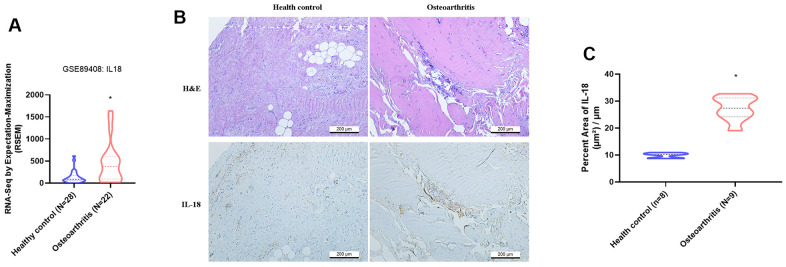
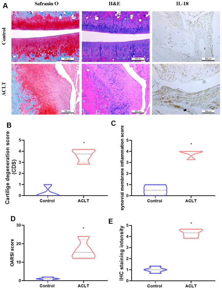
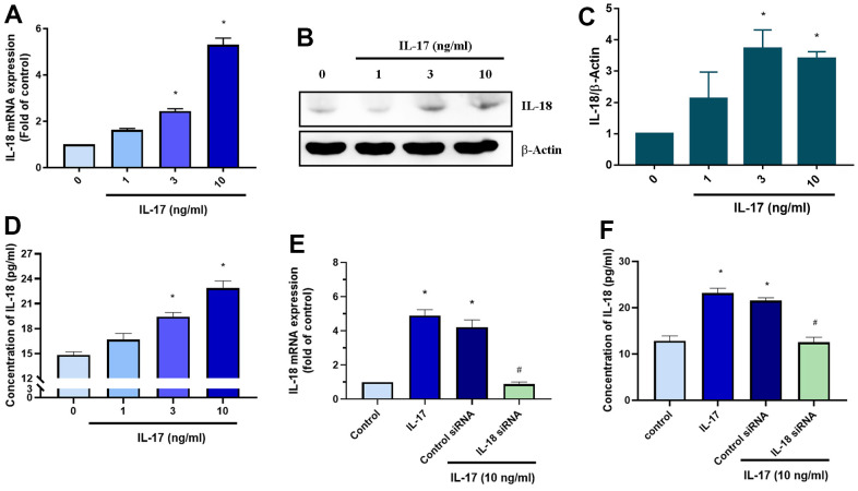
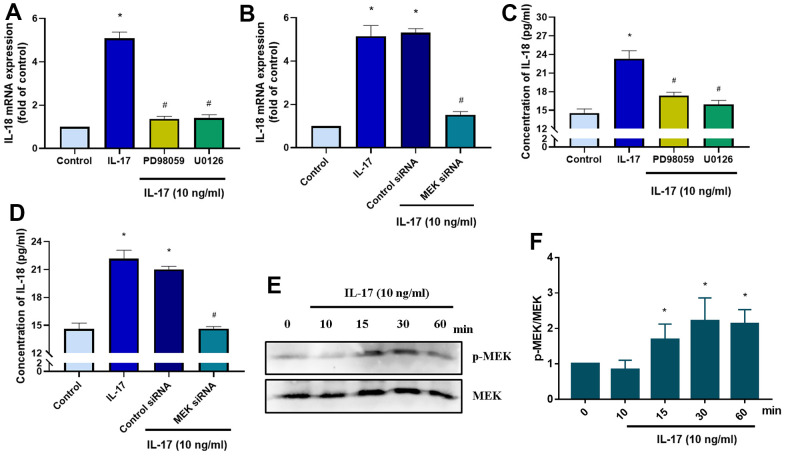
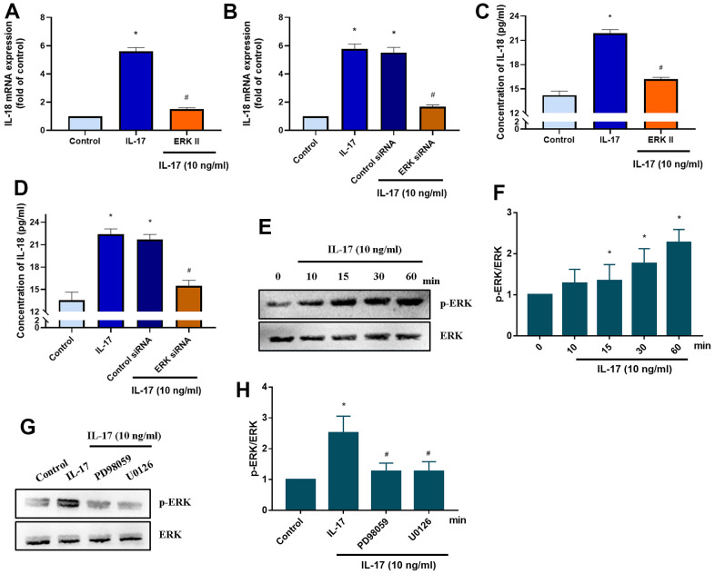
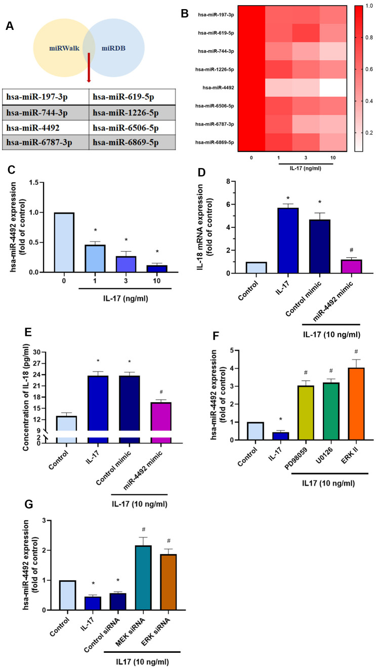
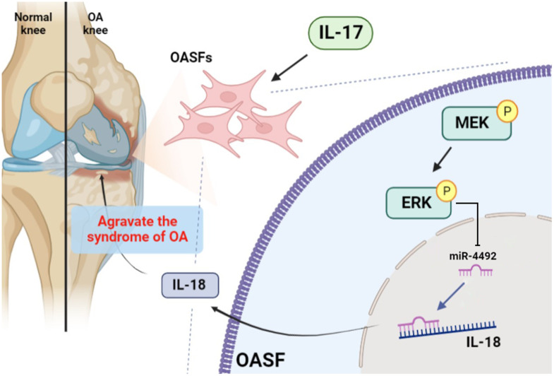
The concept of osteoarthritis (OA) as a low-grade inflammatory joint disorder has been widely accepted. Many inflammatory mediators are implicated in the pathogenesis of OA. Interleukin (IL)-18 is a pleiotropic cytokine with versatile cellular functions that are pathogenetically important in immune responses, as well as autoimmune, inflammatory, and infectious diseases. IL-17, a proinflammatory cytokine mainly secreted by Th17 cells, is upregulated in OA patients. However, the role of IL-17 in OA progression is unclear. The synovial tissues collected from healthy donors and OA patients were used to detect the expression level of IL-18 by IHC stain. The OA synovial fibroblasts (OASFs) were incubated with recombinant IL-17 and subjected to Western blot, qPCR, and ELISA to examine IL-18 expression level. The chemical inhibitors and siRNAs which targeted signal pathways were used to investigate signal pathways involved in IL-17-induced IL-18 expression. The microRNAs which participated IL-18 expression were surveyed with online databases miRWalk and miRDB, followed by validation with qPCR. This study revealed significantly higher levels of IL-18 expression in synovial tissue from OA patients compared with healthy controls, as well as increased IL-18 expression in OASFs from rats with severe OA. In vitro findings indicated that IL-17 dose-dependently promoted IL-18 production in OASFs. Molecular investigations revealed that the MEK/ERK/miR-4492 axis stimulated IL-18 production when OASFs were treated with IL-17. This study provides novel insights into the role of IL-17 in the pathogenesis of OA, which may help to inform OA treatment in the future.

Keywords: IL-17; IL-18; osteoarthritis.


Conflict of interest statement

CONFLICTS OF INTEREST: The authors declare that they have no conflicts of interest.

Figures

Figure 1
Figure 1
IL-18 is upregulated in clinical synovial tissue from OA patients. (A) Patterns of mRNA expression in the GEO dataset GSE890408 in synovial tissue samples from OA patients and normal healthy donors were used to analyze IL-18 expression. (B) Synovial tissues from the study cohorts of OA patients and normal healthy donors were subjected to H&E and IHC staining for detecting IL-18 expression. (C) Quantification of IHC results from Figure 1B. Results are expressed as the means ± S.D. *p<0.05 compared with healthy controls.
Figure 2
Figure 2
Histologic assessments revealed increases in IL-18 expression in ACLT rats. (A) All rats were sacrificed at 8 weeks after surgery. Paraffin-embedded sections of knee joints were subjected to Safranin O/fast green, H&E, and IHC staining for analyses of IL-18 expression. (BD) The severity of knee OA was assessed by cartilage degeneration scores (CDS), synovial membrane inflammation scores, and Osteoarthritis Research Society International (OARSI) scores. (E) Levels of IL-18 expression were evaluated by the intensity of IHC staining. Results are expressed as the means ± S.D. *p<0.05 compared with controls.
Figure 3
Figure 3
IL-17 promotes the production of IL-18 in OASFs. (AD) OASFs were incubated with different concentrations of IL-17 (0–10 ng/mL) for 24 h. Levels of IL-18 expression were detected by qPCR (A), Western blot (B, C), and ELISA (D) analyses. (E, F) OASFs were transfected with IL-18 siRNA then incubated with IL-17 (10 ng/mL) for 24 h and assessed for IL-18 expression. Results are expressed as the means ± S.D. *p<0.05 compared with controls.
Figure 4
Figure 4
IL-17-induced promotion of IL-18 production in OASFs requires MEK signaling. (A) OASFs were pretreated with MEK inhibitors PD98059 (10 μM) or U0126 (5 μM) for 1 h, then incubated with IL-17 (10 ng/mL) for 24 h. IL-18 expression was determined by qPCR. (B) OASFs were transfected with MEK siRNA or control siRNA for 24 h, then stimulated with IL-17 (10 ng/mL) for a further 24 h. IL-18 expression was assessed by qPCR. (C, D) OASFs were treated as described in Figure 4A, 4B. IL-18 production was examined by ELISA. (E) OASFs were stimulated with IL-17 (10 ng/mL) for different time intervals (0–60 min). The total cell lysates were collected and Western blot assessed MEK protein phosphorylation. (F) The quantification result of MEK protein phosphorylation was shown. Results are expressed as the means ± S.D. * p<0.05 compared with controls; # p<0.05 compared with the IL-17-treated group.
Figure 5
Figure 5
ERK signaling mediates IL-18 production in OASFs in response to IL-17 stimulation. (A) OASFs were pretreated with the ERK inhibitor ERKII (5 μM) for 1 h, then incubated with IL-17 (10 ng/mL) for 24 h. IL-18 expression was determined by qPCR. (B) OASFs were transfected with ERK siRNA or control siRNA for 24 h, then stimulated with IL-17 (10 ng/mL) for a further 24 h. IL-18 expression was assessed by qPCR. (C, D) OASFs were treated as described in Figure 5A, 5B. IL-18 production was examined by ELISA. (E, F) OASFs were stimulated with IL-17 (10 ng/mL) for different time intervals (0–60 min). The total cell lysates were collected and Western blot assessed ERK protein phosphorylation. The quantification of blot was shown in Figure 5F. (G, H) OASFs were pretreated with PD98059 (10 μM) or U0126 (5 μM) for 1 h, then stimulated with IL-17 (10 ng/mL) for 60 min. The total cell lysates were collected and Western blot assessed ERK protein phosphorylation. ERK protein was used as the internal control. The quantification of blot was shown in Figure 5H. Results are expressed as the means ± S.D. *p<0.05 compared with controls; #p<0.05 compared with the IL-17-treated group.
Figure 6
Figure 6
The MEK/ERK/miR-4492 axis contributes to IL-18 production in OASFs in response to IL-17 stimulation. (A) Online databases for miRNA target prediction (miRWalk and miRDB) were used to screen for candidate miRNAs that potentially target IL-18 mRNA. (B, C) OASFs were incubated with different concentrations of IL-17 (0–10 ng/mL) for 24 h and miRNA expression was detected by qPCR. (D, E) OASFs were transfected with miR-4492 mimic or control mimic for 24 h, then stimulated with IL-17 (10 ng/mL) for a further 24 h. IL-18 production was assessed by qPCR and ELISA. (F, G) OASFs were pretreated with MER and ERK inhibitor or transfected with MEK, ERK, and control siRNAs, then stimulated with IL-17 (10 ng/mL) for a further 24 h. miR-4492 expression was evaluated by qPCR. Results are expressed as the means ± S.D. * p<0.05 compared with controls; # p<0.05 compared with the IL-17-treated group.
Figure 7
Figure 7
Schematic diagram illustrates the process whereby IL-17 treatment promotes IL-18 production in OASFs. IL-17 treatment upregulates levels of IL-18 expression in OASFs via MEK and ERK signaling and downregulates levels of miR-4492. IL-18 production elicits inflammatory responses during OA progression.

Similar articles

Cited by

References

    1. Cho HJ, Gn KK, Kang JY, Suh KT, Kim TK. Epidemiological characteristics of patellofemoral osteoarthritis in elderly Koreans and its symptomatic contribution in knee osteoarthritis. Knee. 2016; 23:29–34. 10.1016/j.knee.2015.09.003 - DOI - PubMed
    1. Katz JN, Arant KR, Loeser RF. Diagnosis and Treatment of Hip and Knee Osteoarthritis: A Review. JAMA. 2021; 325:568–78. 10.1001/jama.2020.22171 - DOI - PMC - PubMed
    1. Hügle T, Geurts J. What drives osteoarthritis?-synovial versus subchondral bone pathology. Rheumatology (Oxford). 2017; 56:1461–71. 10.1093/rheumatology/kew389 - DOI - PubMed
    1. O’Neill TW, McCabe PS, McBeth J. Update on the epidemiology, risk factors and disease outcomes of osteoarthritis. Best Pract Res Clin Rheumatol. 2018; 32:312–26. 10.1016/j.berh.2018.10.007 - DOI - PubMed
    1. de Liyis BG, Nolan J, Maharjana MA. Fibroblast growth factor receptor 1-bound extracellular vesicle as novel therapy for osteoarthritis. Biomedicine (Taipei). 2022; 12:1–9. 10.37796/2211-8039.1308 - DOI - PMC - PubMed

Publication types