

Minimizing the risk of injury to the popliteal artery during pullout repair of medial meniscus posterior root tears: A cadaveric study
- PMID: 38261907
- PMCID: PMC10797534
- DOI: 10.1016/j.asmart.2023.11.009
Minimizing the risk of injury to the popliteal artery during pullout repair of medial meniscus posterior root tears: A cadaveric study
Abstract
Background: The purpose of this study was to investigate the positional effect of guide pins used in the transtibial pullout repair of medial meniscus posterior root tears on the popliteal artery.
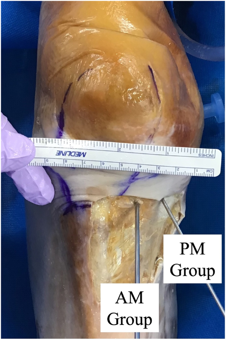
Methods: We used eight cadaveric knees. Two 2.4-mm guide pins were inserted into the posterior root of the medial meniscus at 50° to the articular surface from the medial edge of the tibial tuberosity (anteromedial group) and the anterior edge of the medial collateral ligament (posteromedial group) using an aiming guide placed at the posterior root attachment of the medial meniscus from the anteromedial portal. The posterior capsule was dissected, and the popliteal artery was identified. The positional effect of the guide pins on the popliteal artery was photographed arthroscopically at 0°, 30°, 60°, and 90° knee flexion angles. The popliteal artery diameter and the minimum distance between the popliteal artery center and the guide pin tip were measured.
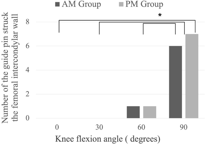
Results: At 90° knee flexion, most of the guide pins in the anteromedial (6 knees; 75 %) and posteromedial groups (7 knees; 87.5 %) collided with the femoral intercondylar wall. The rate of collision was significantly higher at the 90° knee flexion position than that at other angles (p = 0.02). The average shortest distance between the popliteal artery center and the guide pin tip at 0° knee flexion in the posteromedial group (5.4 mm ± 3.4 mm) was significantly greater than that at other knee flexion angles, although the mean distance in the posteromedial group was so negligible that the guide pin could penetrate the popliteal artery.
Conclusions: Knee flexion at 90° causes less damage to the popliteal artery during the transtibial pullout repair of medial meniscus posterior root tears.
Keywords: Medial meniscus posterior root tears; Popliteal artery; Transtibial pullout repair.
© 2023 Asia Pacific Knee, Arthroscopy and Sports Medicine Society. Published by Elsevier (Singapore) Pte Ltd.
Conflict of interest statement
The authors report that they have no conflicts of interest in the authorship and publication of this article.
Figures


Similar articles
-
Transtibial pullout repair of medial meniscus posterior root tear restores physiological rotation of the tibia in the knee-flexed position.Orthop Traumatol Surg Res. 2019 Feb;105(1):113-117. doi: 10.1016/j.otsr.2018.10.005. Epub 2018 Nov 24. Orthop Traumatol Surg Res. 2019. PMID: 30482466
-
Deep flexion helps to avoid popliteal artery injury during all-inside lateral meniscal repair: A cadaveric study.Knee. 2021 Dec;33:159-168. doi: 10.1016/j.knee.2021.09.004. Epub 2021 Oct 6. Knee. 2021. PMID: 34624750
-
The Effect of Knee Flexion Angle on the Neurovascular Safety of All-Inside Lateral Meniscus Repair: A Cadaveric Study.Arthroscopy. 2015 Nov;31(11):2138-44. doi: 10.1016/j.arthro.2015.04.100. Epub 2015 Jul 8. Arthroscopy. 2015. PMID: 26163307
-
Transtibial Pullout Repair Reduces Posterior Extrusion of the Medial Meniscus.Acta Med Okayama. 2019 Dec;73(6):495-501. doi: 10.18926/AMO/57713. Acta Med Okayama. 2019. PMID: 31871331
-
Arthroscopic transtibial pullout repair for posterior meniscus root tears.Oper Orthop Traumatol. 2019 Jun;31(3):248-260. doi: 10.1007/s00064-018-0574-4. Epub 2018 Oct 26. Oper Orthop Traumatol. 2019. PMID: 30367186 Review. English.
References
-
- Bhatia S., LaPrade C.M., Ellman M.B., LaPrade R.F. Meniscal root tears: significance, diagnosis, and treatment. Am J Sports Med. 2014;42(12):3016–3030. - PubMed
-
- Chung K.S., Ha J.K., Ra H.J., Nam G.W., Kim J.G. Pullout fixation of posterior medial meniscus root tears: correlation between meniscus extrusion and midterm clinical results. Am J Sports Med. 2017;45(1):42–49. - PubMed
-
- Aldrich Da R., LoPresti C., Fumich M., Pitluf H., O'Brien W. Pseudoaneurysm Complicating Knee Arthrosc Arthrosc. 1995;11(2):229–230. - PubMed
-
- Alserr A.H., Antonopoulos C.N., Papapetrou A., Kakisis J.D., Brountzos E., Liapis C.D. Endovascular repair of popliteal artery pseudoaneurysm with arteriovenous fistula after knee arthroscopy: case report and literature review. Vasc Endovasc Surg. 2014;48(2):166–170. - PubMed
-
- Thiel W. Die konservierung ganzer leichen in natürlichen farben. Ann Anat Anat Anzeiger. 1992;174(3):185–195. - PubMed
LinkOut - more resources
Full Text Sources
