. 2024 Feb 15;187(4):846-860.e17.
doi: 10.1016/j.cell.2023.12.033. Epub 2024 Jan 22.

Cell surface RNAs control neutrophil recruitment

Affiliations

Cell surface RNAs control neutrophil recruitment

Ningning Zhang et al. Cell. .

Abstract

RNAs localizing to the outer cell surface have been recently identified in mammalian cells, including RNAs with glycan modifications known as glycoRNAs. However, the functional significance of cell surface RNAs and their production are poorly known. We report that cell surface RNAs are critical for neutrophil recruitment and that the mammalian homologs of the sid-1 RNA transporter are required for glycoRNA expression. Cell surface RNAs can be readily detected in murine neutrophils, the elimination of which substantially impairs neutrophil recruitment to inflammatory sites in vivo and reduces neutrophils' adhesion to and migration through endothelial cells. Neutrophil glycoRNAs are predominantly on cell surface, important for neutrophil-endothelial interactions, and can be recognized by P-selectin (Selp). Knockdown of the murine Sidt genes abolishes neutrophil glycoRNAs and functionally mimics the loss of cell surface RNAs. Our data demonstrate the biological importance of cell surface glycoRNAs and highlight a noncanonical dimension of RNA-mediated cellular functions.

Keywords: 45S rRNA; E-selectin; Sidt1; Sidt2; peritonitis; snoRNA; tRNA.


Conflict of interest statement

Declaration of interests The authors declare no competing interests.

Figures

Figure 1.
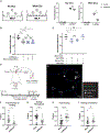
Figure 1.. Detection of RNA on neutrophil surface
(A) HOXB8 cells or differentiated (Diff) HOXB8 cells were treated with or without Ac4ManNAz. RNAs were extracted from the cells, and RNA samples were reacted with DBCO-PEG4-biotin. RNAs were analyzed on an agarose gel (left) and then blotted with an anti-biotin antibody (right). Representative images are shown. (B) Bone marrow (BM) neutrophils were similarly labeled with Ac4ManNAz and analyzed as in (A). After the click-chemistry reaction, RNAs were treated with RNase A in the presence or absence of RNase inhibitor or with Proteinase K (Pro K) or DNase I as indicated. Representative images are shown. (C) The indicated cell types were treated with or without Ac4ManNAz. Cells were then treated extracellularly with RNase A (exRNaseA) or with mock treatment, washed, and then harvested for RNA. Analysis of RNA by gel and blotting was performed similarly as in (A). Representative images are shown. (D) BM neutrophils were treated with or without Ac4ManNAz. Click-chemistry reaction with DBCO-PEG4-biotin were performed directly on live cells without permeabilizing cell membrane. Cells were then treated with exRNaseA or mock conditions. RNAs were harvested and directedly analyzed by gel and blotting without further click-chemistry reactions. Representative images are shown. (E) BM neutrophils were cultured with BrU for 24 h. Cells were further cultured for 30 min with Hoechst. Cells were then treated extracellularly with Proteinase K (exPro K) or with mock treatment and washed. Cells then underwent exRNaseA or mock treatment. Staining by a biotin-conjugated anti-BrU antibody and streptavidin was performed directly on live cells without permeabilizing the cell membrane. Representative images are shown. See also Figure S1.
Figure 2.
Figure 2.. Ablating cell surface RNAs reduces neutrophil recruitment in vivo
(A and B) Bone marrow neutrophils were labeled with a green (CFSE) or a far-red dye. Cells then underwent mock or extracellular RNase A (exRNaseA) treatment before mixing test cells and control cells. Recipient mice were conditioned by intraperitoneal injection (i.p.) of thioglycolate (TG) 1.5 h prior to injecting the mixed neutrophils retro-orbitally (r.o.). Cells were harvested 2.5 h afterward from peripheral blood, bone marrow (BM), spleen, and peritoneum. (A) Schematics of the experiments. (B) Harvested cells were analyzed by flow cytometry, gating on Ly6G+ cells. The ratios between CFSE and far-red-labeled cells were plotted, with each dot representing a recipient mouse. N = 6. Data from a representative experiment are shown. (C) A similarly experiment was performed as in (B), except that test cells were labeled by a far-red dye, whereas control cells were labeled with CFSE. N = 6. Data from a representative experiment are shown. (D) A similar experiment was performed as in (B), except that the mixed neutrophils were directly injected into the peritoneal cavity. In addition, an experimental group of cells treated extracellularly with inactivated RNase A was included. N = 3. Data from a representative experiment are shown. For all panels, error bars represent standard deviation. *p < 0.05; **p < 0.01; ****p < 0.0001; ns: not significant. See also Figure S2 and Table S1.
Figure 3.
Figure 3.. Ablating cell surface RNAs reduces neutrophil-endothelial cell interaction
(A) Bone marrow neutrophils underwent mock (control) or extracellular RNase A (exRNaseA) treatment. Cell migration was analyzed in a transwell assay with one group of experiments having endothelial cells (ECs) plated to cover the top surface of the transwell insert, whereas another group without ECs. For testing migration, neutrophils were added to the top chamber with the chemoattractant fMLP (2 mM) added to the bottom chamber. Left: schematics for experiments. Right: the numbers of the migrated cells were quantified and normalized as percentages of input cells. Each dot represents a biological replicate. N = 3. Data from representative experiments are shown. (B) The static adhesion of bone marrow neutrophils to ECs was analyzed, where ECs were pre-plated as a confluent layer in culture dishes. Neutrophils were dye-labeled (to facilitate counting on ECs) and treated with or without exRNaseA. In some conditions, glycoRNAs purified from neutrophils were used to pre-block the ECs. For assaying adhesion, neutrophils were added to EC-plated dishes for 10 min, followed by washes to remove unattached or loosely attached neutrophils. Cells were then counted under microscope, with adherence quantified as the number of neutrophils averaged across at least 10 random imaging fields, and normalized to the control (no exRNaseA, no glycoRNA blocking) group. Each dot represents a biological replicate. N = 6. Data from a representative experiment are shown. (C) A similar experiment as in (B) was performed, except that in addition to blocking ECs with glycoRNAs, blocking with the glycan fraction and the RNA fraction of the neutrophil glycoRNAs was performed. N = 4. Data from a representative experiment are shown. (D–I) Intravital imaging experiments were performed to assay differential neutrophil-endothelial interactions by mock and exRNaseA-treated neutrophils in vivo. (D) The schematics of the experiment. A 1:1 mixture of mock and exRNaseA-treated neutrophils was administered via intracarotid (i.c.) injection to C57BL/6 mice previously stimulated with TNF-α. Intravital confocal microscopy was performed to analyze rolling and adhesion of injected neutrophils to the blood vessels of the cremaster muscle. exRNaseA- or mock-treated neutrophils were labeled with CFSE or far-red cell tracer. Two independent experiments for each labeling combination were carried out. (E) A representative confocal image of the cremaster microcirculation, with blood vessel linings indicated by blue lines, with different states of neutrophils indicated by symbols in the legend. (F–I) The numbers of neutrophils that were free rolling, transitioned from free rolling to rolling, kept rolling throughout the imagining interval, or transitioned from rolling to stable adhesion were quantified. Data were normalized to reflect the fractions of neutrophils within a given color that display the corresponding behavior in a field. Each dot represents data from a field at one of the imaging intervals. For all panels, except for (F)–(I), error bars represent standard deviations. For (F)–(I), error bars represent SEM. *p < 0.05; ***p < 0.001; ****p < 0.0001; ns: not significant. See also Figure S3 and Video S1.
Figure 4.
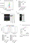
Figure 4.. Neutrophilic glycoRNAs interact with Selp
(A) Endothelial cells (ECs) were blocked with anti-Selp or anti-Sele antibodies before biotin-labeled glycoRNAs were added to assay binding to ECs. GlycoRNAs were purified from Ac4ManNAz-treated bone marrow neutrophils and labeled with biotin through click chemistry. ECs were dissociated by an enzyme-free buffer, and the levels of glycoRNA binding were quantified using flow cytometry via streptavidin. ECs analyzed without streptavidin (Ctrl) or without antibody blocking were used as controls. Representative flow cytometry plots are shown. (B) Data in (A) were quantified as mean fluorescence intensity, normalized by the control condition without blocking antibody. Each dot represents a biological replicate. N = 3. Data from a representative experiment are shown. (C) RNAs were harvested from bone marrow neutrophils treated with or without Ac4ManNAz. RNAs from cells with Ac4ManNAz were labeled with biotin through click chemistry. RNAs were analyzed by gel and were blotted with anti-biotin, or with recombinant proteins of Selp-Fc fusion or Sele-Fc fusion. Representative images are shown. (D) A similar experiment as in (C) was performed, including a condition with purified RNAs treated with RNase A before gel and blot analysis. (E) Bone marrow (BM) neutrophils underwent exRNaseA or mock treatment. Recombinant Selp-Fc or Sele-Fc were used to bind to live neutrophils. Top: representative flow cytometry plots showing the levels of Selp and Sezdxle binding signals, with unstained cells as negative control (Ctrl). Bottom: quantified mean fluorescence intensity was normalized by the mock treatment condition. N = 3. Data from a representative experiment are shown. (F) BM neutrophils were treated with mock or exRNaseA and subjected to in vitro adhesion assay to WT and Selp KO ECs. N = 3. Data from a representative experiment are shown. For all panels, error bars represent standard deviation. ***p < 0.001; ****p < 0.0001; ns: not significant. See also Figure S4.
Figure 5.
Figure 5.. Sidt family transporters are required for glycoRNA production and neutrophil-endothelial cell interaction
(A) Model 1 depicts that RNA (red) molecules are released from originating cells and captured by other cells. Model 2 depicts cell-intrinsic production and transfer of RNA to cell surface. Experiments were designed to distinguish the two models via the schematics on the right. BM neutrophils were treated with or without Ac4ManNAz for 24 h, with Ac4ManNAz-treated cells labeled with a green dye and untreated cells with a red dye. Cells were mixed together and co-cultured for 24–72 h before FACS to isolate live green and red cells. RNAs were harvested from the sorted cell populations and analyzed by gel and blotted for glycoRNA similar to experiments in Figure 1A. (B) Data for the experiment in (A), with representative images shown. (C) Cas9-expressing HOXB8 cells were transduced with a control sgRNA (wild type or WT) or two independent sets of sgRNAs to knock down (KD) the expression of Sidt1 and Sidt2. Cells were differentiated toward neutrophils and treated with Ac4ManNAz. Cells were further subjected to exRNAseA or mock treatment. RNAs were harvested and subjected to analysis of glycoRNA via gel and immunoblotting. Representative images are shown. (D) Neutrophils differentiated from WT and Sidt-KD HOXB8 cells were subjected to exRNaseA or mock treatment. Cells were analyzed for adhesion to endothelial cells (ECs) similar to Figure 3B. Each dot represents a biological replicate. N = 6. Data from a representative experiment are shown. (E) Cells in (D) were analyzed for transmigration with or without ECs, similar to experiments in Figure 3A. Each dot represents a biological replicate. N = 3. Data from a representative experiment are shown. (F) WT and Sidt-KD HOXB8 cells were differentiated in vivo to obtain WT and Sidt-KD neutrophils. These neutrophils were dye-labeled, mixed, and injected into recipient mice to assay for recruitment to the peritoneum in the acute peritonitis model in Figure 2A. Ratios of KD to WT cells were quantified in the indicated tissues, with data from two independent sgRNA KD vectors. N = 3. Data from representative experiments are shown. For all panels, error bars represent standard deviation. **p < 0.01; ***p < 0.001; ****p < 0.0001; ns: not significant. See also Figure S5.
Figure 6.
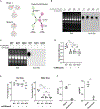
Figure 6.. Murine neutrophil glycoRNAs are primarily small RNAs from noncoding transcripts
(A) Total RNAs from BM neutrophils (input) were purified for glycoRNAs by WGA beads. The purified glycoRNA fraction was digested with PNGase F. The sizes of RNAs were analyzed on a Bioanalyzer with a broad range high sensitivity RNA analysis cartridge. Numbers indicate lengths of nucleotides. Of note, the exact sizes of small RNAs may not be fully accurate on this analysis platform. (B) Purification and processing schemes for small RNA library preparation. HOXB8 cells, HOXB8-derived neutrophils, and BM neutrophils were used. (C) Top: sequences enriched in the glycoRNA fractions from the indicated samples were counted and categorized based on the types of RNAs that they mapped to. Bottom: the total abundance of enriched glycoRNA sequences that mapped to the indicated RNA categories was calculated for each of the samples. RPM, reads per million mapped reads. (D) The levels of RNA sequences in purified glycoRNA samples were plotted against those of the input samples, with the cell types indicated at the top. The red dashed boxes indicate sequences that are more enriched and show positive correlation to input sequence abundance, whereas the green dashed boxes indicate sequences that are depleted from the purified glycoRNA samples. See also Figure S6 and Table S2.

References

    1. Huang N, Fan X, Zaleta-Rivera K, Nguyen TC, Zhou J, Luo Y, Gao J, Fang RH, Yan Z, Chen ZB, et al. (2020). Natural display of nuclear-encoded RNA on the cell surface and its impact on cell interaction. Genome Biol 21, 225. - PMC - PubMed
    1. Flynn RA, Pedram K, Malaker SA, Batista PJ, Smith BAH, Johnson AG, George BM, Majzoub K, Villalta PW, Carette JE, et al. (2021). Small RNAs are modified with N-glycans and displayed on the surface of living cells. Cell 184, 3109–3124.e22. - PMC - PubMed
    1. Wang GG, Calvo KR, Pasillas MP, Sykes DB, Häcker H, and Kamps MP (2006). Quantitative production of macrophages or neutrophils ex vivo using conditional Hoxb8. Nat. Methods 3, 287–293. - PubMed
    1. Orosz A, Walzog B, and Mócsai A (2021). In vivo functions of mouse neutrophils derived from HoxB8-transduced conditionally immortalized myeloid progenitors. J. Immunol 206, 432–445. - PubMed
    1. McDonald JU, Cortini A, Rosas M, Fossati-Jimack L, Ling GS, Lewis KJ, Dewitt S, Liddiard K, Brown GD, Jones SA, et al. (2011). In vivo functional analysis and genetic modification of in vitro -derived mouse neutrophils. FASEB J 25, 1972–1982. - PubMed